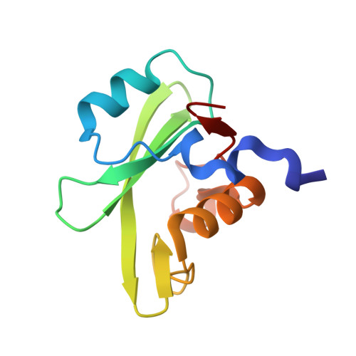
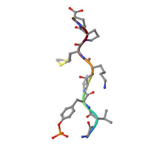
7CIO

Molecular interactions of cytoplasmic region of CTLA-4 with SH2 domains of PI3-kinase


Experimental Data Snapshot

  • Method: X-RAY DIFFRACTION
  • Resolution: 1.10 Å
  • R-Value Free: 0.179 
  • R-Value Work: 0.154 
  • R-Value Observed: 0.155 

wwPDB Validation   3D Report Full Report


This is version 1.2 of the entry. See complete history


Literature

Molecular interactions of the CTLA-4 cytoplasmic region with the phosphoinositide 3-kinase SH2 domains.

Iiyama, M.Numoto, N.Ogawa, S.Kuroda, M.Morii, H.Abe, R.Ito, N.Oda, M.

(2021) Mol Immunol 131: 51-59

  • DOI: https://doi.org/10.1016/j.molimm.2020.12.002
  • Primary Citation of Related Structures:  
    7CIO

  • PubMed Abstract: 

    During T-cell regulation, T-cell receptors and CD28 lead to signaling activation, while T-lymphocyte antigen 4 (CTLA-4) is known to lead to downregulation, similar to programmed cell death-1 (PD-1). In the cytoplasmic tails of CD28 and CTLA-4, phosphoinositide 3-kinase (PI3K) binds to the consensus sequence including phosphotyrosine via SH2 domains, N- and C-terminal SH2 domains (nSH2 and cSH2), of its regulatory subunit, p85. In this study, we determined the crystal structure of a CTLA-4-derived phosphopeptide in complex with a Cys-substituted mutant of cSH2, C656S/C659V/C670L, at a 1.1 Å resolution. Phosphotyrosine of the bound peptide is tightly accommodated by the residues Arg631, Arg649, Ser651, and Ser652, similar to the cSH2 wild-type recognition mode of CD28, as reported previously. Upon the Cys mutation, the cSH2 thermal stability increased while the CTLA-4 binding affinity slightly changed. The binding experiments also showed that the binding affinity of CTLA-4 by cSH2 was approximately two orders of magnitude lower than that of CD28. Similar to CD28 binding, the CTLA-4 binding affinity of nSH2 was lower than that of cSH2. The complex structure of nSH2 and CTLA-4 was modeled, and compared with the crystal structure of cSH2 mutant and CTLA-4. The difference in the binding affinity between CD28 and CTLA-4, along with the difference between nSH2 and cSH2, could be explained by the 3D structures, which would be closely correlated with the respective T-cell signaling.


  • Organizational Affiliation

    Graduate School of Life and Environmental Sciences, Kyoto Prefectural University, 1-5 Hangi-cho, Shimogamo, Sakyo-ku, Kyoto 606-8522, Japan.


Macromolecules
Find similar proteins by:  (by identity cutoff)  |  3D Structure
Entity ID: 1
MoleculeChains Sequence LengthOrganismDetailsImage
Phosphatidylinositol 3-kinase regulatory subunit alpha109Homo sapiensMutation(s): 3 
Gene Names: PIK3R1GRB1
UniProt & NIH Common Fund Data Resources
Find proteins for P27986 (Homo sapiens)
Explore P27986 
Go to UniProtKB:  P27986
PHAROS:  P27986
GTEx:  ENSG00000145675 
Entity Groups  
Sequence Clusters30% Identity50% Identity70% Identity90% Identity95% Identity100% Identity
UniProt GroupP27986
Sequence Annotations
Expand
  • Reference Sequence

Find similar proteins by:  Sequence   |   3D Structure  

Entity ID: 2
MoleculeChains Sequence LengthOrganismDetailsImage
Cytotoxic T-lymphocyte protein 48Homo sapiensMutation(s): 0 
UniProt & NIH Common Fund Data Resources
Find proteins for P16410 (Homo sapiens)
Explore P16410 
Go to UniProtKB:  P16410
PHAROS:  P16410
GTEx:  ENSG00000163599 
Entity Groups  
UniProt GroupP16410
Sequence Annotations
Expand
  • Reference Sequence
Small Molecules
Modified Residues  1 Unique
IDChains TypeFormula2D DiagramParent
PTR
Query on PTR
B
L-PEPTIDE LINKINGC9 H12 N O6 PTYR
Experimental Data & Validation

Experimental Data

  • Method: X-RAY DIFFRACTION
  • Resolution: 1.10 Å
  • R-Value Free: 0.179 
  • R-Value Work: 0.154 
  • R-Value Observed: 0.155 
  • Space Group: P 21 21 21
Unit Cell:
Length ( Å )Angle ( ˚ )
a = 41.221α = 90
b = 41.563β = 90
c = 59.628γ = 90
Software Package:
Software NamePurpose
PHENIXrefinement
XDSdata reduction
XDSdata scaling
PHASERphasing

Structure Validation

View Full Validation Report


Entry History 

Deposition Data

Revision History  (Full details and data files)

  • Version 1.0: 2020-12-09
    Type: Initial release
  • Version 1.1: 2021-03-17
    Changes: Database references
  • Version 1.2: 2023-11-29
    Changes: Data collection, Database references, Refinement description