7D27

Structure of UDP-N-acetylmuramoyl-L-alanyl-D-glutamate--2, 6-diaminopimelate ligase


Experimental Data Snapshot

  • Method: X-RAY DIFFRACTION
  • Resolution: 2.48 Å
  • R-Value Free: 0.260 
  • R-Value Work: 0.228 

wwPDB Validation   3D Report Full Report


This is version 1.1 of the entry. See complete history


Literature

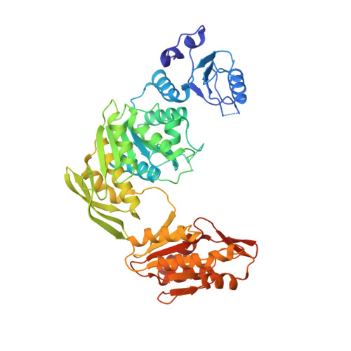
Wide-open conformation of UDP-MurNc-tripeptide ligase revealed by the substrate-free structure of MurE from Acinetobacter baumannii.

Jung, K.H.Kim, Y.G.Kim, C.M.Ha, H.J.Lee, C.S.Lee, J.H.Park, H.H.

(2021) FEBS Lett 595: 275-283

  • DOI: https://doi.org/10.1002/1873-3468.14007
  • Primary Citation of Related Structures:  
    7D27

  • PubMed Abstract: 

    MurE ligase catalyzes the attachment of meso-diaminopimelic acid to the UDP-MurNAc- l -Ala- d -Glu using ATP and producing UDP-MurNAc- l -Ala- d -Glu-meso-A 2 pm during bacterial cell wall biosynthesis. Owing to the critical role of this enzyme, MurE is considered an attractive target for antibacterial drugs. Despite extensive studies on MurE ligase, the structural dynamics of its conformational changes are still elusive. In this study, we present the substrate-free structure of MurE from Acinetobacter baumannii, which is an antibiotic-resistant superbacterium that has threatened global public health. The structure revealed that MurE has a wide-open conformation and undergoes wide-open, intermediately closed, and fully closed dynamic conformational transition. Unveiling structural dynamics of MurE will help to understand the working mechanism of this ligase and to design next-generation antibiotics targeting MurE.


  • Organizational Affiliation

    Department of Global Innovative Drugs, Graduate School of Chung-Ang University, Seoul, Korea.


Macromolecules
Find similar proteins by:  (by identity cutoff)  |  3D Structure
Entity ID: 1
MoleculeChains Sequence LengthOrganismDetailsImage
UDP-N-acetylmuramoyl-L-alanyl-D-glutamate--2,6-diaminopimelate ligase499Acinetobacter baumanniiMutation(s): 0 
Gene Names: murEEA667_001595EA685_00960EA746_009110
EC: 6.3.2.13
Entity Groups  
Sequence Clusters30% Identity50% Identity70% Identity90% Identity95% Identity100% Identity
Sequence Annotations
Expand
  • Reference Sequence
Experimental Data & Validation

Experimental Data

  • Method: X-RAY DIFFRACTION
  • Resolution: 2.48 Å
  • R-Value Free: 0.260 
  • R-Value Work: 0.228 
  • Space Group: P 42 21 2
Unit Cell:
Length ( Å )Angle ( ˚ )
a = 119.96α = 90
b = 119.96β = 90
c = 116.73γ = 90
Software Package:
Software NamePurpose
REFMACrefinement
MxDCdata collection
XDSdata scaling
PHENIXphasing

Structure Validation

View Full Validation Report


Entry History & Funding Information

Deposition Data


Funding OrganizationLocationGrant Number
National Research Foundation (NRF, Korea)Korea, Republic Of--

Revision History  (Full details and data files)

  • Version 1.0: 2021-07-28
    Type: Initial release
  • Version 1.1: 2023-11-29
    Changes: Data collection, Database references, Derived calculations, Refinement description