


crystal structure of type III polyketide synthase from Mycobacterium marinum
Pratap, S., Kant, A., Saxena, P., Krishnan, V.To be published.
Experimental Data Snapshot
Starting Model: experimental
View more details
Entity ID: 1 | |||||
|---|---|---|---|---|---|
| Molecule | Chains | Sequence Length | Organism | Details | Image |
| Chalcone synthase-like protein | A [auth B] | 413 | Mycobacterium marinum M | Mutation(s): 0 Gene Names: MMAR_2190 | 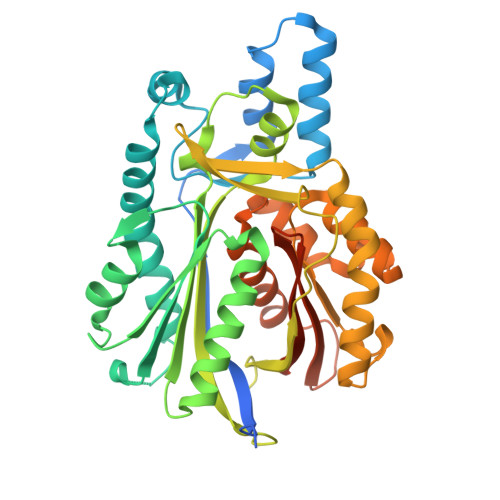 |
UniProt | |||||
Entity Groups | |||||
| Sequence Clusters | 30% Identity50% Identity70% Identity90% Identity95% Identity100% Identity | ||||
| UniProt Group | B2HNE1 | ||||
Sequence AnnotationsExpand | |||||
Reference Sequence | |||||
Entity ID: 2 | |||||
|---|---|---|---|---|---|
| Molecule | Chains | Sequence Length | Organism | Details | Image |
| Chalcone/stilbene synthase | B [auth A] | 413 | Mycobacterium marinum M | Mutation(s): 0 Gene Names: MMAR_2190 | 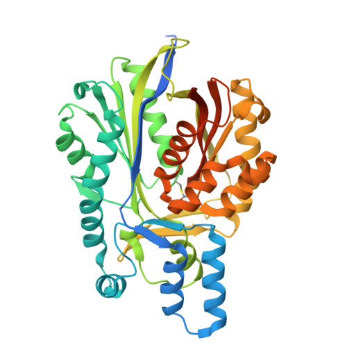 |
UniProt | |||||
Entity Groups | |||||
| Sequence Clusters | 30% Identity50% Identity70% Identity90% Identity95% Identity100% Identity | ||||
| UniProt Group | B2HNE1 | ||||
Sequence AnnotationsExpand | |||||
Reference Sequence | |||||
| Ligands 1 Unique | |||||
|---|---|---|---|---|---|
| ID | Chains | Name / Formula / InChI Key | 2D Diagram | 3D Interactions | |
| OCA (Subject of Investigation/LOI) Download:Ideal Coordinates CCD File | C [auth B], D [auth A] | OCTANOIC ACID (CAPRYLIC ACID) C8 H16 O2 WWZKQHOCKIZLMA-UHFFFAOYSA-N |  | ||
| Modified Residues 1 Unique | |||||
|---|---|---|---|---|---|
| ID | Chains | Type | Formula | 2D Diagram | Parent |
| MCS Query on MCS | B [auth A] | L-PEPTIDE LINKING | C6 H9 N O5 S |  | CYS |
| Length ( Å ) | Angle ( ˚ ) |
|---|---|
| a = 55.732 | α = 90 |
| b = 99.518 | β = 90 |
| c = 126.707 | γ = 90 |
| Software Name | Purpose |
|---|---|
| REFMAC | refinement |
| XDS | data reduction |
| STARANISO | data scaling |
| SHELXDE | phasing |
| Funding Organization | Location | Grant Number |
|---|---|---|
| Department of Science & Technology (DST, India) | India | CRG/2018/002229 |


