


Interaction of metals with PfGrx1
Manickam, Y., Sharma, A.To be published.
Experimental Data Snapshot
Entity ID: 1 | |||||
|---|---|---|---|---|---|
| Molecule | Chains | Sequence Length | Organism | Details | Image |
| Glutaredoxin | 111 | Plasmodium falciparum 3D7 | Mutation(s): 0 Gene Names: PF3D7_0306300 EC: 1.8.4.2 | 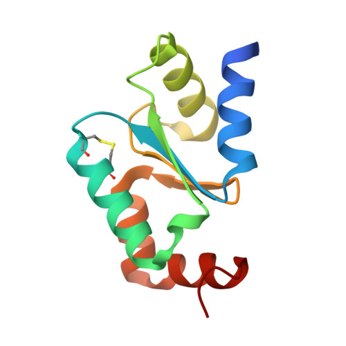 | |
UniProt | |||||
Entity Groups | |||||
| Sequence Clusters | 30% Identity50% Identity70% Identity90% Identity95% Identity100% Identity | ||||
| UniProt Group | Q9NLB2 | ||||
Sequence AnnotationsExpand | |||||
Reference Sequence | |||||
| Ligands 3 Unique | |||||
|---|---|---|---|---|---|
| ID | Chains | Name / Formula / InChI Key | 2D Diagram | 3D Interactions | |
| MPO Download:Ideal Coordinates CCD File | B [auth A] | 3[N-MORPHOLINO]PROPANE SULFONIC ACID C7 H15 N O4 S DVLFYONBTKHTER-UHFFFAOYSA-N |  | ||
| PT Download:Ideal Coordinates CCD File | D [auth A], E [auth A] | PLATINUM (II) ION Pt HRGDZIGMBDGFTC-UHFFFAOYSA-N |  | ||
| MPD (Subject of Investigation/LOI) Download:Ideal Coordinates CCD File | C [auth A] | (4S)-2-METHYL-2,4-PENTANEDIOL C6 H14 O2 SVTBMSDMJJWYQN-YFKPBYRVSA-N |  | ||
| Length ( Å ) | Angle ( ˚ ) |
|---|---|
| a = 48.492 | α = 90 |
| b = 48.492 | β = 90 |
| c = 82.69 | γ = 120 |
| Software Name | Purpose |
|---|---|
| HKL-2000 | data scaling |
| PHENIX | refinement |
| PDB_EXTRACT | data extraction |
| HKL-2000 | data reduction |
| Auto-Rickshaw | phasing |
| SHELXDE | phasing |
| Coot | model building |
| Funding Organization | Location | Grant Number |
|---|---|---|
| Department of Biotechnology (DBT, India) | India | -- |


