Download Mendeley
ApmA Is a Unique Aminoglycoside Antibiotic Acetyltransferase That Inactivates Apramycin.
Bordeleau, E., Stogios, P.J., Evdokimova, E., Koteva, K., Savchenko, A., Wright, G.D.(2021) mBio 12
- PubMed: 33563840 Search on PubMedSearch on PubMed Central
- DOI: https://doi.org/10.1128/mBio.02705-20
- Primary Citation Related Structures:
7JM0, 7JM1 - PubMed Abstract:
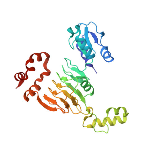
Apramycin is an aminoglycoside antibiotic with the potential to be developed to combat multidrug-resistant pathogens. Its unique structure evades the clinically widespread mechanisms of aminoglycoside resistance that currently compromise the efficacy of other members in this drug class. Of the aminoglycoside-modifying enzymes that chemically alter these antibiotics, only AAC(3)-IVa has been demonstrated to confer resistance to apramycin through N -acetylation. Knowledge of other modification mechanisms is important to successfully develop apramycin for clinical use. Here, we show that ApmA is structurally unique among the previously described aminoglycoside-modifying enzymes and capable of conferring a high level of resistance to apramycin. In vitro experiments indicated ApmA to be an N -acetyltransferase, but in contrast to AAC(3)-IVa, ApmA has a unique regiospecificity of the acetyl transfer to the N2' position of apramycin. Crystallographic analysis of ApmA conclusively showed that this enzyme is an acetyltransferase from the left-handed β-helix protein superfamily (LβH) with a conserved active site architecture. The success of apramycin will be dependent on consideration of the impact of this potential form of clinical resistance. IMPORTANCE Apramycin is an aminoglycoside antibiotic that has been traditionally used in veterinary medicine. Recently, it has become an attractive candidate to repurpose in the fight against multidrug-resistant pathogens prioritized by the World Health Organization. Its atypical structure circumvents most of the clinically relevant mechanisms of resistance that impact this class of antibiotics. Prior to repurposing apramycin, it is important to understand the resistance mechanisms that could be a liability. Our study characterizes the most recently identified apramycin resistance element, apmA We show ApmA does not belong to the protein families typically associated with aminoglycoside resistance and is responsible for modifying a different site on the molecule. The data presented will be critical in the development of apramycin derivatives that will evade apmA in the event it becomes prevalent in the clinic.
- David Braley Centre for Antibiotics Discovery, M.G. DeGroote Institute for Infectious Disease Research, Department of Biochemistry and Biomedical Sciences, McMaster University, Hamilton, Ontario, Canada.
Organizational Affiliation: