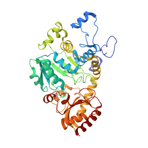
7KB1

Complex of O-acety-L-homoserine aminocarboxypropyltransferase (MetY) from Thermotoga maritima and a key reaction intermediate


Experimental Data Snapshot

  • Method: X-RAY DIFFRACTION
  • Resolution: 1.85 Å
  • R-Value Free: 0.262 
  • R-Value Work: 0.227 
  • R-Value Observed: 0.229 

Starting Model: experimental
View more details

wwPDB Validation   3D Report Full Report


Ligand Structure Quality Assessment 


This is version 1.3 of the entry. See complete history


Literature

Structures and kinetics of Thermotoga maritima MetY reveal new insights into the predominant sulfurylation enzyme of bacterial methionine biosynthesis.

Brewster, J.L.Pachl, P.McKellar, J.L.O.Selmer, M.Squire, C.J.Patrick, W.M.

(2021) J Biol Chem 296: 100797-100797

  • DOI: https://doi.org/10.1016/j.jbc.2021.100797
  • Primary Citation of Related Structures:  
    7KB0, 7KB1

  • PubMed Abstract: 

    Bacterial methionine biosynthesis can take place by either the trans-sulfurylation route or direct sulfurylation. The enzymes responsible for trans-sulfurylation have been characterized extensively because they occur in model organisms such as Escherichia coli. However, direct sulfurylation is actually the predominant route for methionine biosynthesis across the phylogenetic tree. In this pathway, most bacteria use an O-acetylhomoserine aminocarboxypropyltransferase (MetY) to catalyze the formation of homocysteine from O-acetylhomoserine and bisulfide. Despite the widespread distribution of MetY, this pyridoxal 5'-phosphate-dependent enzyme remains comparatively understudied. To address this knowledge gap, we have characterized the MetY from Thermotoga maritima (TmMetY). At its optimal temperature of 70 °C, TmMetY has a turnover number (apparent k cat  = 900 s -1 ) that is 10- to 700-fold higher than the three other MetY enzymes for which data are available. We also present crystal structures of TmMetY in the internal aldimine form and, fortuitously, with a β,γ-unsaturated ketimine reaction intermediate. This intermediate is identical to that found in the catalytic cycle of cystathionine γ-synthase (MetB), which is a homologous enzyme from the trans-sulfurylation pathway. By comparing the TmMetY and MetB structures, we have identified Arg270 as a critical determinant of specificity. It helps to wall off the active site of TmMetY, disfavoring the binding of the first MetB substrate, O-succinylhomoserine. It also ensures a strict specificity for bisulfide as the second substrate of MetY by occluding the larger MetB substrate, cysteine. Overall, this work illuminates the subtle structural mechanisms by which homologous pyridoxal 5'-phosphate-dependent enzymes can effect different catalytic, and therefore metabolic, outcomes.


  • Organizational Affiliation

    Department of Biochemistry, University of Otago, Dunedin, New Zealand.


Macromolecules
Find similar proteins by:  (by identity cutoff)  |  3D Structure
Entity ID: 1
MoleculeChains Sequence LengthOrganismDetailsImage
O-acetyl-L-homoserine sulfhydrylase
A, B, C, D
430Thermotoga maritima MSB8Mutation(s): 0 
Gene Names: TM_0882Tmari_0884
EC: 2.5.1
UniProt
Find proteins for Q9WZY4 (Thermotoga maritima (strain ATCC 43589 / DSM 3109 / JCM 10099 / NBRC 100826 / MSB8))
Explore Q9WZY4 
Go to UniProtKB:  Q9WZY4
Entity Groups  
Sequence Clusters30% Identity50% Identity70% Identity90% Identity95% Identity100% Identity
UniProt GroupQ9WZY4
Sequence Annotations
Expand
  • Reference Sequence
Small Molecules
Ligands 5 Unique
IDChains Name / Formula / InChI Key2D Diagram3D Interactions
WBJ (Subject of Investigation/LOI)
Query on WBJ

Download Ideal Coordinates CCD File 
E [auth A],
G [auth B],
I [auth C]
(2E)-2-[({3-hydroxy-2-methyl-5-[(phosphonooxy)methyl]pyridin-4-yl}methyl)imino]but-3-enoic acid
C12 H15 N2 O7 P
ILCWREDNKMRIGC-GXDHUFHOSA-N
PLP (Subject of Investigation/LOI)
Query on PLP

Download Ideal Coordinates CCD File 
J [auth D]PYRIDOXAL-5'-PHOSPHATE
C8 H10 N O6 P
NGVDGCNFYWLIFO-UHFFFAOYSA-N
PGE
Query on PGE

Download Ideal Coordinates CCD File 
H [auth B]TRIETHYLENE GLYCOL
C6 H14 O4
ZIBGPFATKBEMQZ-UHFFFAOYSA-N
MG
Query on MG

Download Ideal Coordinates CCD File 
K [auth D]MAGNESIUM ION
Mg
JLVVSXFLKOJNIY-UHFFFAOYSA-N
NA
Query on NA

Download Ideal Coordinates CCD File 
F [auth A]SODIUM ION
Na
FKNQFGJONOIPTF-UHFFFAOYSA-N
Experimental Data & Validation

Experimental Data

  • Method: X-RAY DIFFRACTION
  • Resolution: 1.85 Å
  • R-Value Free: 0.262 
  • R-Value Work: 0.227 
  • R-Value Observed: 0.229 
  • Space Group: P 1 21 1
Unit Cell:
Length ( Å )Angle ( ˚ )
a = 67.58α = 90
b = 151.34β = 112.041
c = 91.39γ = 90
Software Package:
Software NamePurpose
PHENIXrefinement
XDSdata reduction
XSCALEdata scaling
MOLREPphasing

Structure Validation

View Full Validation Report


Ligand Structure Quality Assessment 


Entry History & Funding Information

Deposition Data


Funding OrganizationLocationGrant Number
Marsden FundNew Zealand--

Revision History  (Full details and data files)

  • Version 1.0: 2021-06-23
    Type: Initial release
  • Version 1.1: 2021-07-28
    Changes: Database references
  • Version 1.2: 2024-03-06
    Changes: Data collection, Database references, Refinement description
  • Version 1.3: 2024-04-03
    Changes: Refinement description