7KML | pdb_00007kml

cryo-EM structure of SARS-CoV-2 spike in complex with Fab 15033-7, three RBDs bound


Experimental Data Snapshot

  • Method: ELECTRON MICROSCOPY
  • Resolution: 3.80 Å
  • Aggregation State: PARTICLE 
  • Reconstruction Method: SINGLE PARTICLE 

Starting Models: experimental
View more details

wwPDB Validation 3D Report Full Report

Validation slider image for 7KML

This is version 1.3 of the entry. See complete history

Literature

Tetravalent SARS-CoV-2 Neutralizing Antibodies Show Enhanced Potency and Resistance to Escape Mutations.

Miersch, S.Li, Z.Saberianfar, R.Ustav, M.Brett Case, J.Blazer, L.Chen, C.Ye, W.Pavlenco, A.Gorelik, M.Garcia Perez, J.Subramania, S.Singh, S.Ploder, L.Ganaie, S.Chen, R.E.Leung, D.W.Pandolfi, P.P.Novelli, G.Matusali, G.Colavita, F.Capobianchi, M.R.Jain, S.Gupta, J.B.Amarasinghe, G.K.Diamond, M.S.Rini, J.Sidhu, S.S.

(2021) J Mol Biology 433: 167177-167177

  • DOI: https://doi.org/10.1016/j.jmb.2021.167177
  • Primary Citation Related Structures: 
    7KLG, 7KLH, 7KMK, 7KML, 7KXJ, 7KXK

  • PubMed Abstract: 

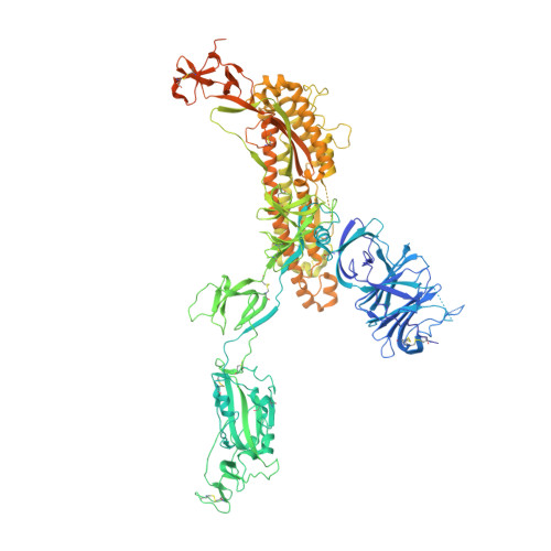
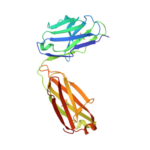
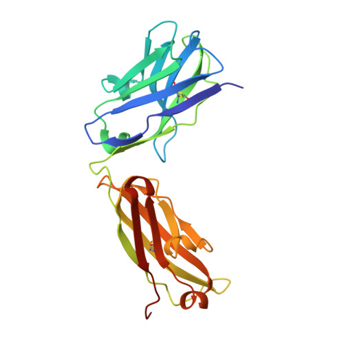
    Neutralizing antibodies (nAbs) hold promise as therapeutics against COVID-19. Here, we describe protein engineering and modular design principles that have led to the development of synthetic bivalent and tetravalent nAbs against SARS-CoV-2. The best nAb targets the host receptor binding site of the viral S-protein and tetravalent versions block entry with a potency exceeding bivalent nAbs by an order of magnitude. Structural studies show that both the bivalent and tetravalent nAbs can make multivalent interactions with a single S-protein trimer, consistent with the avidity and potency of these molecules. Significantly, we show that the tetravalent nAbs show increased tolerance to potential virus escape mutants and an emerging variant of concern. Bivalent and tetravalent nAbs can be produced at large-scale and are as stable and specific as approved antibody drugs. Our results provide a general framework for enhancing antiviral therapies against COVID-19 and related viral threats, and our strategy can be applied to virtually any antibody drug.


  • Organizational Affiliation
    • The Donnelly Centre, University of Toronto, Toronto, Canada.

Macromolecule Content 

  • Total Structure Weight: 580.56 kDa 
  • Atom Count: 35,136 
  • Modeled Residue Count: 4,413 
  • Deposited Residue Count: 5,139 
  • Unique protein chains: 3

Macromolecules

Find similar proteins by:|  3D Structure
Entity ID: 1
MoleculeChains  Sequence LengthOrganismDetailsImage
Spike glycoprotein
A, B, C
1,274Severe acute respiratory syndrome coronavirus 2Mutation(s): 0 
Gene Names: S2
UniProt
Find proteins for P0DTC2 (Severe acute respiratory syndrome coronavirus 2)
Explore P0DTC2 
Go to UniProtKB:  P0DTC2
Entity Groups
Sequence Clusters30% Identity50% Identity70% Identity90% Identity95% Identity100% Identity
UniProt GroupP0DTC2
Glycosylation
Glycosylation Sites: 15Go to GlyGen: P0DTC2-1
Sequence Annotations
Expand
Reference Sequence
Find similar proteins by:|  3D Structure
Entity ID: 2
MoleculeChains  Sequence LengthOrganismDetailsImage
Fab 15033-7 light chainD [auth L],
F [auth M],
H [auth N]
214Homo sapiensMutation(s): 0 
Find similar proteins by:|  3D Structure
Entity ID: 3
MoleculeChains  Sequence LengthOrganismDetailsImage
Fab 15033-7 heavy chainE [auth H],
G [auth I],
I [auth J]
225Homo sapiensMutation(s): 0 

Oligosaccharides

Help  
Entity ID: 5
MoleculeChains Length2D Diagram GlycosylationD Interactions
beta-D-mannopyranose-(1-4)-2-acetamido-2-deoxy-beta-D-glucopyranose-(1-4)-2-acetamido-2-deoxy-beta-D-glucopyranose
DA [auth d],
EA [auth e],
L [auth F],
M [auth G],
U,
DA [auth d],
EA [auth e],
L [auth F],
M [auth G],
U,
V
3N-Glycosylation
Glycosylation Resources
GlyTouCan: G15407YE
GlyCosmos: G15407YE
GlyGen: G15407YE

Small Molecules

Ligands 1 Unique
IDChains Name / Formula / InChI Key2D Diagram3D Interactions
NAG

Query on NAG


Download:Ideal Coordinates CCD File
AB [auth C]
BB [auth C]
KA [auth A]
LA [auth A]
MA [auth A]
AB [auth C],
BB [auth C],
KA [auth A],
LA [auth A],
MA [auth A],
NA [auth A],
OA [auth A],
PA [auth A],
QA [auth B],
RA [auth B],
SA [auth B],
TA [auth B],
UA [auth B],
VA [auth B],
WA [auth C],
XA [auth C],
YA [auth C],
ZA [auth C]
2-acetamido-2-deoxy-beta-D-glucopyranose
C8 H15 N O6
OVRNDRQMDRJTHS-FMDGEEDCSA-N

Experimental Data & Validation

Experimental Data

  • Method: ELECTRON MICROSCOPY
  • Resolution: 3.80 Å
  • Aggregation State: PARTICLE 
  • Reconstruction Method: SINGLE PARTICLE 
EM Software:
TaskSoftware PackageVersion
RECONSTRUCTIONcryoSPARC2.15
MODEL REFINEMENTRosettaEM
MODEL REFINEMENTPHENIX

Structure Validation

View Full Validation Report


Entry History 

& Funding Information

Deposition Data


Funding OrganizationLocationGrant Number
Canadian Institutes of Health Research (CIHR)Canada--

Revision History  (Full details and data files)

  • Version 1.0: 2021-02-10
    Type: Initial release
  • Version 1.1: 2021-08-18
    Changes: Database references
  • Version 1.2: 2021-08-25
    Changes: Database references
  • Version 1.3: 2024-11-13
    Changes: Data collection, Database references, Refinement description, Structure summary