7L36

PEPCK steady-state structure with Mn and GTP


Experimental Data Snapshot

  • Method: X-RAY DIFFRACTION
  • Resolution: 1.84 Å
  • R-Value Free: 0.199 
  • R-Value Work: 0.152 
  • R-Value Observed: 0.155 

wwPDB Validation   3D Report Full Report


Ligand Structure Quality Assessment 


This is version 1.1 of the entry. See complete history


Literature

Millisecond mix-and-quench crystallography (MMQX) enables time-resolved studies of PEPCK with remote data collection.

Clinger, J.A.Moreau, D.W.McLeod, M.J.Holyoak, T.Thorne, R.E.

(2021) IUCrJ 8: 784-792

  • DOI: https://doi.org/10.1107/S2052252521007053
  • Primary Citation of Related Structures:  
    7L36, 7L3M, 7L3V

  • PubMed Abstract: 

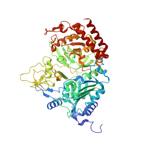
    Time-resolved crystallography of biomolecules in action has advanced rapidly as methods for serial crystallography have improved, but the large number of crystals and the complex experimental infrastructure that are required remain serious obstacles to its widespread application. Here, millisecond mix-and-quench crystallography (MMQX) has been developed, which yields millisecond time-resolved data using far fewer crystals and routine remote synchrotron data collection. To demonstrate the capabilities of MMQX, the conversion of oxaloacetic acid to phosphoenolpyruvate by phosphoenolpyruvate carboxy-kinase (PEPCK) is observed with a time resolution of 40 ms. By lowering the entry barrier to time-resolved crystallography, MMQX should enable a broad expansion in structural studies of protein dynamics.


  • Organizational Affiliation

    Physics Department, Cornell University, 142 Sciences Drive, Ithaca, NY 14853, USA.


Macromolecules
Find similar proteins by:  (by identity cutoff)  |  3D Structure
Entity ID: 1
MoleculeChains Sequence LengthOrganismDetailsImage
Phosphoenolpyruvate carboxykinase, cytosolic [GTP]624Rattus norvegicusMutation(s): 0 
Gene Names: Pck1
EC: 4.1.1.32 (PDB Primary Data), 2.7.11 (PDB Primary Data)
UniProt
Find proteins for P07379 (Rattus norvegicus)
Explore P07379 
Go to UniProtKB:  P07379
Entity Groups  
Sequence Clusters30% Identity50% Identity70% Identity90% Identity95% Identity100% Identity
UniProt GroupP07379
Sequence Annotations
Expand
  • Reference Sequence
Experimental Data & Validation

Experimental Data

  • Method: X-RAY DIFFRACTION
  • Resolution: 1.84 Å
  • R-Value Free: 0.199 
  • R-Value Work: 0.152 
  • R-Value Observed: 0.155 
  • Space Group: P 1 21 1
Unit Cell:
Length ( Å )Angle ( ˚ )
a = 44.38α = 90
b = 118.55β = 109.53
c = 60.233γ = 90
Software Package:
Software NamePurpose
PHENIXrefinement
Aimlessdata scaling
PDB_EXTRACTdata extraction
DIALSdata reduction

Structure Validation

View Full Validation Report


Ligand Structure Quality Assessment 


Entry History & Funding Information

Deposition Data


Funding OrganizationLocationGrant Number
National Institutes of Health/National Institute of General Medical Sciences (NIH/NIGMS)United States1R01GM127528-01
Natural Sciences and Engineering Research Council (NSERC, Canada)Canada--

Revision History  (Full details and data files)

  • Version 1.0: 2021-10-13
    Type: Initial release
  • Version 1.1: 2023-10-18
    Changes: Data collection, Refinement description