7LA4

Integrin AlphaIIbBeta3-PT25-2 Complex


Experimental Data Snapshot

  • Method: ELECTRON MICROSCOPY
  • Resolution: 3.30 Å
  • Aggregation State: PARTICLE 
  • Reconstruction Method: SINGLE PARTICLE 

wwPDB Validation   3D Report Full Report


This is version 1.1 of the entry. See complete history


Literature

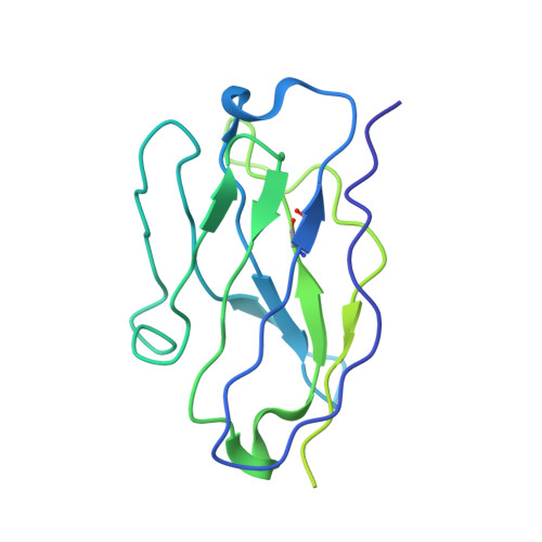
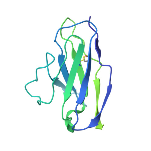
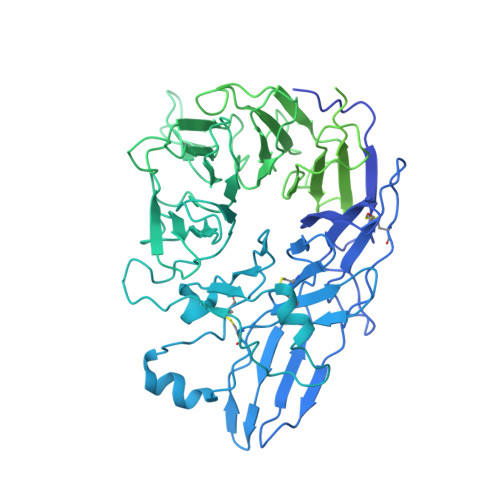
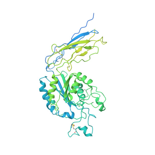
Electron microscopy shows that binding of monoclonal antibody PT25-2 primes integrin alpha IIb beta 3 for ligand binding.

Nesic, D.Bush, M.Spasic, A.Li, J.Kamata, T.Handa, M.Filizola, M.Walz, T.Coller, B.S.

(2021) Blood Adv 5: 1781-1790

  • DOI: https://doi.org/10.1182/bloodadvances.2020004166
  • Primary Citation of Related Structures:  
    7LA4

  • PubMed Abstract: 

    The murine monoclonal antibody (mAb) PT25-2 induces αIIbβ3 to bind ligand and initiate platelet aggregation. The underlying mechanism is unclear, because previous mutagenesis studies suggested that PT25-2 binds to the αIIb β propeller, a site distant from the Arg-Gly-Asp-binding pocket. To elucidate the mechanism, we studied the αIIbβ3-PT25-2 Fab complex by negative-stain and cryo-electron microscopy (EM). We found that PT25-2 binding results in αIIbβ3 partially exposing multiple ligand-induced binding site epitopes and adopting extended conformations without swing-out of the β3 hybrid domain. The cryo-EM structure showed PT25-2 binding to the αIIb residues identified by mutagenesis but also to 2 additional regions. Overlay of the cryo-EM structure with the bent αIIbβ3 crystal structure showed that binding of PT25-2 creates clashes with the αIIb calf-1/calf-2 domains, suggesting that PT25-2 selectively binds to partially or fully extended receptor conformations and prevents a return to its bent conformation. Kinetic studies of the binding of PT25-2 compared with mAbs 10E5 and 7E3 support this hypothesis. We conclude that PT25-2 induces αIIbβ3 ligand binding by binding to extended conformations and by preventing the interactions between the αIIb and β3 leg domains and subsequently the βI and β3 leg domains required for the bent-closed conformation.


  • Organizational Affiliation

    Laboratory of Blood and Vascular Biology and.


Macromolecules
Find similar proteins by:  (by identity cutoff)  |  3D Structure
Entity ID: 1
MoleculeChains Sequence LengthOrganismDetailsImage
PT25-2 heavy chainA [auth H]214Mus musculusMutation(s): 0 
Entity Groups  
Sequence Clusters30% Identity50% Identity70% Identity90% Identity95% Identity100% Identity
Sequence Annotations
Expand
  • Reference Sequence
Find similar proteins by:  (by identity cutoff)  |  3D Structure
Entity ID: 2
MoleculeChains Sequence LengthOrganismDetailsImage
PT25-2 light chainB [auth L]214Mus musculusMutation(s): 0 
Entity Groups  
Sequence Clusters30% Identity50% Identity70% Identity90% Identity95% Identity100% Identity
Sequence Annotations
Expand
  • Reference Sequence
Find similar proteins by:  (by identity cutoff)  |  3D Structure
Entity ID: 3
MoleculeChains Sequence LengthOrganismDetailsImage
Integrin alpha-IIbC [auth A]1,039Homo sapiensMutation(s): 0 
Gene Names: ITGA2BGP2BITGAB
UniProt & NIH Common Fund Data Resources
Find proteins for P08514 (Homo sapiens)
Explore P08514 
Go to UniProtKB:  P08514
PHAROS:  P08514
GTEx:  ENSG00000005961 
Entity Groups  
Sequence Clusters30% Identity50% Identity70% Identity90% Identity95% Identity100% Identity
UniProt GroupP08514
Sequence Annotations
Expand
  • Reference Sequence
Find similar proteins by:  (by identity cutoff)  |  3D Structure
Entity ID: 4
MoleculeChains Sequence LengthOrganismDetailsImage
Integrin beta-3D [auth B]788Homo sapiensMutation(s): 0 
Gene Names: ITGB3GP3A
UniProt & NIH Common Fund Data Resources
Find proteins for P05106 (Homo sapiens)
Explore P05106 
Go to UniProtKB:  P05106
PHAROS:  P05106
GTEx:  ENSG00000259207 
Entity Groups  
Sequence Clusters30% Identity50% Identity70% Identity90% Identity95% Identity100% Identity
UniProt GroupP05106
Sequence Annotations
Expand
  • Reference Sequence
Oligosaccharides

Help

Entity ID: 5
MoleculeChains Length2D Diagram Glycosylation3D Interactions
2-acetamido-2-deoxy-beta-D-glucopyranose-(1-4)-2-acetamido-2-deoxy-beta-D-glucopyranoseE [auth C]2N/AN-Glycosylation
Glycosylation Resources
GlyTouCan:  G42666HT
GlyCosmos:  G42666HT
GlyGen:  G42666HT
Entity ID: 6
MoleculeChains Length2D Diagram Glycosylation3D Interactions
alpha-D-mannopyranose-(1-3)-alpha-D-mannopyranose-(1-4)-2-acetamido-2-deoxy-beta-D-glucopyranose-(1-4)-2-acetamido-2-deoxy-beta-D-glucopyranoseF [auth D]4N/AN-Glycosylation
Glycosylation Resources
GlyTouCan:  G48068RF
GlyCosmos:  G48068RF
GlyGen:  G48068RF
Entity ID: 7
MoleculeChains Length2D Diagram Glycosylation3D Interactions
alpha-D-mannopyranose-(1-4)-2-acetamido-2-deoxy-beta-D-glucopyranose-(1-4)-2-acetamido-2-deoxy-beta-D-glucopyranoseG [auth E]3N/AN-Glycosylation
Glycosylation Resources
GlyTouCan:  G62182OO
GlyCosmos:  G62182OO
GlyGen:  G62182OO
Small Molecules
Ligands 3 Unique
IDChains Name / Formula / InChI Key2D Diagram3D Interactions
NAG
Query on NAG

Download Ideal Coordinates CCD File 
L [auth A]2-acetamido-2-deoxy-beta-D-glucopyranose
C8 H15 N O6
OVRNDRQMDRJTHS-FMDGEEDCSA-N
CA
Query on CA

Download Ideal Coordinates CCD File 
H [auth A]
I [auth A]
J [auth A]
K [auth A]
N [auth B]
H [auth A],
I [auth A],
J [auth A],
K [auth A],
N [auth B],
O [auth B]
CALCIUM ION
Ca
BHPQYMZQTOCNFJ-UHFFFAOYSA-N
MG
Query on MG

Download Ideal Coordinates CCD File 
M [auth B]MAGNESIUM ION
Mg
JLVVSXFLKOJNIY-UHFFFAOYSA-N
Experimental Data & Validation

Experimental Data

  • Method: ELECTRON MICROSCOPY
  • Resolution: 3.30 Å
  • Aggregation State: PARTICLE 
  • Reconstruction Method: SINGLE PARTICLE 

Structure Validation

View Full Validation Report


Entry History & Funding Information

Deposition Data


Funding OrganizationLocationGrant Number
National Institutes of Health/National Heart, Lung, and Blood Institute (NIH/NHLBI)United States2R01HL019278-43

Revision History  (Full details and data files)

  • Version 1.0: 2022-01-12
    Type: Initial release
  • Version 1.1: 2022-07-27
    Changes: Database references