7LAA | pdb_00007laa

Structure of SARS-CoV-2 S protein in complex with Receptor Binding Domain antibody DH1041


Experimental Data Snapshot

  • Method: ELECTRON MICROSCOPY
  • Resolution: 3.42 Å
  • Aggregation State: PARTICLE 
  • Reconstruction Method: SINGLE PARTICLE 

Starting Model: experimental
View more details

wwPDB Validation 3D Report Full Report

Validation slider image for 7LAA

This is version 1.5 of the entry. See complete history

Literature

In vitro and in vivo functions of SARS-CoV-2 infection-enhancing and neutralizing antibodies.

Li, D.Edwards, R.J.Manne, K.Martinez, D.R.Schafer, A.Alam, S.M.Wiehe, K.Lu, X.Parks, R.Sutherland, L.L.Oguin 3rd, T.H.McDanal, C.Perez, L.G.Mansouri, K.Gobeil, S.M.C.Janowska, K.Stalls, V.Kopp, M.Cai, F.Lee, E.Foulger, A.Hernandez, G.E.Sanzone, A.Tilahun, K.Jiang, C.Tse, L.V.Bock, K.W.Minai, M.Nagata, B.M.Cronin, K.Gee-Lai, V.Deyton, M.Barr, M.Von Holle, T.Macintyre, A.N.Stover, E.Feldman, J.Hauser, B.M.Caradonna, T.M.Scobey, T.D.Rountree, W.Wang, Y.Moody, M.A.Cain, D.W.DeMarco, C.T.Denny, T.N.Woods, C.W.Petzold, E.W.Schmidt, A.G.Teng, I.T.Zhou, T.Kwong, P.D.Mascola, J.R.Graham, B.S.Moore, I.N.Seder, R.Andersen, H.Lewis, M.G.Montefiori, D.C.Sempowski, G.D.Baric, R.S.Acharya, P.Haynes, B.F.Saunders, K.O.

(2021) Cell 184: 4203

  • DOI: https://doi.org/10.1016/j.cell.2021.06.021
  • Primary Citation Related Structures: 
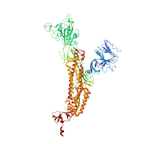
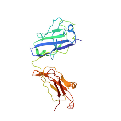
    7LAA, 7LAB, 7LCN, 7LD1

  • PubMed Abstract: 

    SARS-CoV-2-neutralizing antibodies (NAbs) protect against COVID-19. A concern regarding SARS-CoV-2 antibodies is whether they mediate disease enhancement. Here, we isolated NAbs against the receptor-binding domain (RBD) or the N-terminal domain (NTD) of SARS-CoV-2 spike from individuals with acute or convalescent SARS-CoV-2 or a history of SARS-CoV infection. Cryo-electron microscopy of RBD and NTD antibodies demonstrated function-specific modes of binding. Select RBD NAbs also demonstrated Fc receptor-γ (FcγR)-mediated enhancement of virus infection in vitro, while five non-neutralizing NTD antibodies mediated FcγR-independent in vitro infection enhancement. However, both types of infection-enhancing antibodies protected from SARS-CoV-2 replication in monkeys and mice. Three of 46 monkeys infused with enhancing antibodies had higher lung inflammation scores compared to controls. One monkey had alveolar edema and elevated bronchoalveolar lavage inflammatory cytokines. Thus, while in vitro antibody-enhanced infection does not necessarily herald enhanced infection in vivo, increased lung inflammation can rarely occur in SARS-CoV-2 antibody-infused macaques.


  • Organizational Affiliation
    • Duke Human Vaccine Institute, Duke University School of Medicine, Durham, NC 27710, USA; Department of Medicine, Duke University School of Medicine, Durham, NC 27710, USA.

Macromolecule Content 

  • Total Structure Weight: 431.49 kDa 
  • Atom Count: 23,712 
  • Modeled Residue Count: 3,002 
  • Deposited Residue Count: 3,806 
  • Unique protein chains: 3

Macromolecules

Find similar proteins by:|  3D Structure
Entity ID: 1
MoleculeChains  Sequence LengthOrganismDetailsImage
Spike glycoproteinA,
B,
E [auth C]
1,121Severe acute respiratory syndrome coronavirus 2Mutation(s): 0 
Gene Names: S2
UniProt
Find proteins for P0DTC2 (Severe acute respiratory syndrome coronavirus 2)
Explore P0DTC2 
Go to UniProtKB:  P0DTC2
Entity Groups
Sequence Clusters30% Identity50% Identity70% Identity90% Identity95% Identity100% Identity
UniProt GroupP0DTC2
Glycosylation
Glycosylation Sites: 11Go to GlyGen: P0DTC2-1
Sequence Annotations
Expand
Reference Sequence
Find similar proteins by:|  3D Structure
Entity ID: 2
MoleculeChains  Sequence LengthOrganismDetailsImage
DH1041 heavy chainC [auth H]226Homo sapiensMutation(s): 0 
Find similar proteins by:|  3D Structure
Entity ID: 3
MoleculeChains  Sequence LengthOrganismDetailsImage
DH1041 light chainD [auth L]217Homo sapiensMutation(s): 0 

Oligosaccharides

Help  
Entity ID: 4
MoleculeChains Length2D Diagram GlycosylationD Interactions
2-acetamido-2-deoxy-beta-D-glucopyranose-(1-4)-2-acetamido-2-deoxy-beta-D-glucopyranose
F [auth D],
G [auth E],
H [auth F],
I [auth G],
J [auth I],
F [auth D],
G [auth E],
H [auth F],
I [auth G],
J [auth I],
M,
O,
Q,
R,
T,
V
2N-Glycosylation
Glycosylation Resources
GlyTouCan: G42666HT
GlyCosmos: G42666HT
GlyGen: G42666HT
Entity ID: 5
MoleculeChains Length2D Diagram GlycosylationD Interactions
beta-D-mannopyranose-(1-4)-2-acetamido-2-deoxy-beta-D-glucopyranose-(1-4)-2-acetamido-2-deoxy-beta-D-glucopyranose
K [auth J],
L [auth K],
N,
P,
S,
K [auth J],
L [auth K],
N,
P,
S,
U
3N-Glycosylation
Glycosylation Resources
GlyTouCan: G15407YE
GlyCosmos: G15407YE
GlyGen: G15407YE

Small Molecules

Ligands 1 Unique
IDChains Name / Formula / InChI Key2D Diagram3D Interactions
NAG

Query on NAG


Download:Ideal Coordinates CCD File
AA [auth A]
BA [auth B]
CA [auth B]
DA [auth B]
EA [auth B]
AA [auth A],
BA [auth B],
CA [auth B],
DA [auth B],
EA [auth B],
FA [auth B],
GA [auth B],
HA [auth C],
IA [auth C],
JA [auth C],
KA [auth C],
LA [auth C],
MA [auth C],
W [auth A],
X [auth A],
Y [auth A],
Z [auth A]
2-acetamido-2-deoxy-beta-D-glucopyranose
C8 H15 N O6
OVRNDRQMDRJTHS-FMDGEEDCSA-N

Experimental Data & Validation

Experimental Data

  • Method: ELECTRON MICROSCOPY
  • Resolution: 3.42 Å
  • Aggregation State: PARTICLE 
  • Reconstruction Method: SINGLE PARTICLE 
EM Software:
TaskSoftware PackageVersion
MODEL REFINEMENTISOLDE
MODEL REFINEMENTPHENIX
MODEL REFINEMENTCoot
RECONSTRUCTIONcryoSPARC

Structure Validation

View Full Validation Report


Entry History 

& Funding Information

Deposition Data


Funding OrganizationLocationGrant Number
National Institutes of Health/National Institute Of Allergy and Infectious Diseases (NIH/NIAID)United StatesAI145687

Revision History  (Full details and data files)

  • Version 1.0: 2021-03-17
    Type: Initial release
  • Version 1.1: 2021-03-24
    Changes: Database references
  • Version 1.2: 2021-07-07
    Changes: Data collection, Database references
  • Version 1.3: 2021-07-28
    Changes: Database references
  • Version 1.4: 2021-08-18
    Changes: Database references
  • Version 1.5: 2024-11-20
    Changes: Data collection, Database references, Refinement description, Structure summary