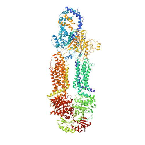
7LKP

Structure of ATP-free human ABCA4


Experimental Data Snapshot

  • Method: ELECTRON MICROSCOPY
  • Resolution: 3.27 Å
  • Aggregation State: PARTICLE 
  • Reconstruction Method: SINGLE PARTICLE 

wwPDB Validation   3D Report Full Report


This is version 2.1 of the entry. See complete history


Literature

Molecular structures of the eukaryotic retinal importer ABCA4.

Liu, F.Lee, J.Chen, J.

(2021) Elife 10

  • DOI: https://doi.org/10.7554/eLife.63524
  • Primary Citation of Related Structures:  
    7LKP, 7LKZ

  • PubMed Abstract: 

    The ATP-binding cassette (ABC) transporter family contains thousands of members with diverse functions. Movement of the substrate, powered by ATP hydrolysis, can be outward (export) or inward (import). ABCA4 is a eukaryotic importer transporting retinal to the cytosol to enter the visual cycle. It also removes toxic retinoids from the disc lumen. Mutations in ABCA4 cause impaired vision or blindness. Despite decades of clinical, biochemical, and animal model studies, the molecular mechanism of ABCA4 is unknown. Here, we report the structures of human ABCA4 in two conformations. In the absence of ATP, ABCA4 adopts an outward-facing conformation, poised to recruit substrate. The presence of ATP induces large conformational changes that could lead to substrate release. These structures provide a molecular basis to understand many disease-causing mutations and a rational guide for new experiments to uncover how ABCA4 recruits, flips, and releases retinoids.


  • Organizational Affiliation

    Laboratory of Membrane Biology and Biophysics, The Rockefeller University, New York, United States.


Macromolecules
Find similar proteins by:  (by identity cutoff)  |  3D Structure
Entity ID: 1
MoleculeChains Sequence LengthOrganismDetailsImage
Retinal-specific phospholipid-transporting ATPase ABCA42,273Homo sapiensMutation(s): 0 
Gene Names: ABCA4ABCR
EC: 7.6.2.1
Membrane Entity: Yes 
UniProt & NIH Common Fund Data Resources
Find proteins for P78363 (Homo sapiens)
Explore P78363 
Go to UniProtKB:  P78363
PHAROS:  P78363
GTEx:  ENSG00000198691 
Entity Groups  
Sequence Clusters30% Identity50% Identity70% Identity90% Identity95% Identity100% Identity
UniProt GroupP78363
Glycosylation
Glycosylation Sites: 8Go to GlyGen: P78363-1
Sequence Annotations
Expand
  • Reference Sequence
Oligosaccharides

Help

Entity ID: 2
MoleculeChains Length2D Diagram Glycosylation3D Interactions
beta-D-mannopyranose-(1-4)-2-acetamido-2-deoxy-beta-D-glucopyranose-(1-4)-2-acetamido-2-deoxy-beta-D-glucopyranose
B
3N-Glycosylation
Glycosylation Resources
GlyTouCan:  G15407YE
GlyCosmos:  G15407YE
GlyGen:  G15407YE
Entity ID: 3
MoleculeChains Length2D Diagram Glycosylation3D Interactions
beta-D-mannopyranose-(1-3)-[beta-D-mannopyranose-(1-6)]beta-D-mannopyranose-(1-4)-2-acetamido-2-deoxy-beta-D-glucopyranose-(1-4)-2-acetamido-2-deoxy-beta-D-glucopyranose
C
5N-Glycosylation
Glycosylation Resources
GlyTouCan:  G89225LT
GlyCosmos:  G89225LT
GlyGen:  G89225LT
Small Molecules
Ligands 4 Unique
IDChains Name / Formula / InChI Key2D Diagram3D Interactions
AJP
Query on AJP

Download Ideal Coordinates CCD File 
M [auth A]Digitonin
C56 H92 O29
UVYVLBIGDKGWPX-KUAJCENISA-N
POV
Query on POV

Download Ideal Coordinates CCD File 
K [auth A]
S [auth A]
T [auth A]
U [auth A]
V [auth A]
K [auth A],
S [auth A],
T [auth A],
U [auth A],
V [auth A],
W [auth A],
X [auth A]
(2S)-3-(hexadecanoyloxy)-2-[(9Z)-octadec-9-enoyloxy]propyl 2-(trimethylammonio)ethyl phosphate
C42 H82 N O8 P
WTJKGGKOPKCXLL-PFDVCBLKSA-N
CLR
Query on CLR

Download Ideal Coordinates CCD File 
J [auth A]
L [auth A]
N [auth A]
O [auth A]
P [auth A]
J [auth A],
L [auth A],
N [auth A],
O [auth A],
P [auth A],
Q [auth A],
R [auth A]
CHOLESTEROL
C27 H46 O
HVYWMOMLDIMFJA-DPAQBDIFSA-N
NAG
Query on NAG

Download Ideal Coordinates CCD File 
D [auth A]
E [auth A]
F [auth A]
G [auth A]
H [auth A]
D [auth A],
E [auth A],
F [auth A],
G [auth A],
H [auth A],
I [auth A]
2-acetamido-2-deoxy-beta-D-glucopyranose
C8 H15 N O6
OVRNDRQMDRJTHS-FMDGEEDCSA-N
Experimental Data & Validation

Experimental Data

  • Method: ELECTRON MICROSCOPY
  • Resolution: 3.27 Å
  • Aggregation State: PARTICLE 
  • Reconstruction Method: SINGLE PARTICLE 

Structure Validation

View Full Validation Report


Entry History & Funding Information

Deposition Data


Funding OrganizationLocationGrant Number
Howard Hughes Medical Institute (HHMI)United States--

Revision History  (Full details and data files)

  • Version 1.0: 2021-03-03
    Type: Initial release
  • Version 2.0: 2021-08-25
    Changes: Advisory, Atomic model, Data collection, Database references
  • Version 2.1: 2024-11-06
    Changes: Data collection, Structure summary