7NE1

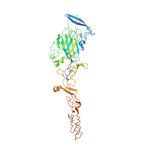
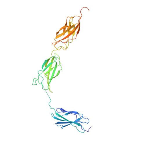
Structure of the complex between Netrin-1 and its receptor Neogenin


Experimental Data Snapshot

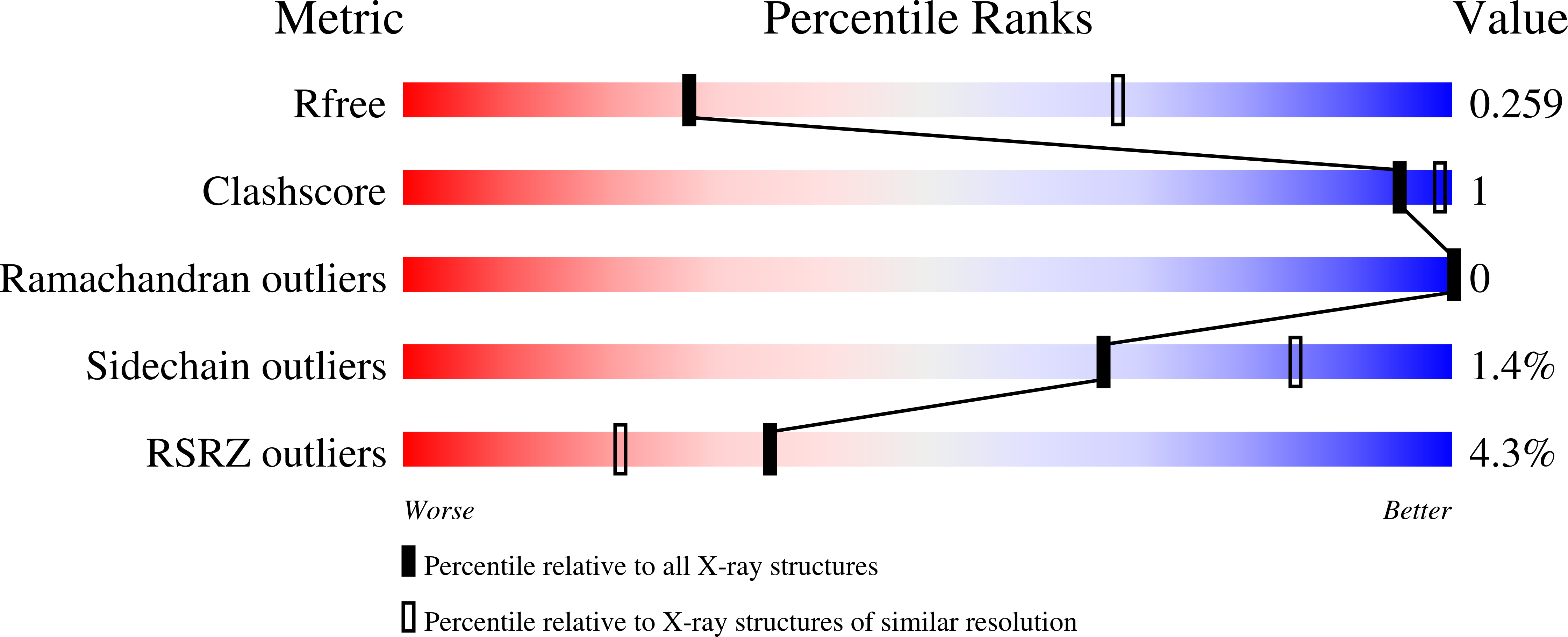
  • Method: X-RAY DIFFRACTION
  • Resolution: 3.15 Å
  • R-Value Free: 0.239 
  • R-Value Work: 0.212 
  • R-Value Observed: 0.214 

Starting Models: experimental
View more details

wwPDB Validation   3D Report Full Report


Ligand Structure Quality Assessment 


This is version 1.3 of the entry. See complete history


Literature

Simultaneous binding of Guidance Cues NET1 and RGM blocks extracellular NEO1 signaling.

Robinson, R.A.Griffiths, S.C.van de Haar, L.L.Malinauskas, T.van Battum, E.Y.Zelina, P.Schwab, R.A.Karia, D.Malinauskaite, L.Brignani, S.van den Munkhof, M.H.Dudukcu, O.De Ruiter, A.A.Van den Heuvel, D.M.A.Bishop, B.Elegheert, J.Aricescu, A.R.Pasterkamp, R.J.Siebold, C.

(2021) Cell 184: 2103

  • DOI: https://doi.org/10.1016/j.cell.2021.02.045
  • Primary Citation of Related Structures:  
    7NDG, 7NE0, 7NE1

  • PubMed Abstract: 

    During cell migration or differentiation, cell surface receptors are simultaneously exposed to different ligands. However, it is often unclear how these extracellular signals are integrated. Neogenin (NEO1) acts as an attractive guidance receptor when the Netrin-1 (NET1) ligand binds, but it mediates repulsion via repulsive guidance molecule (RGM) ligands. Here, we show that signal integration occurs through the formation of a ternary NEO1-NET1-RGM complex, which triggers reciprocal silencing of downstream signaling. Our NEO1-NET1-RGM structures reveal a "trimer-of-trimers" super-assembly, which exists in the cell membrane. Super-assembly formation results in inhibition of RGMA-NEO1-mediated growth cone collapse and RGMA- or NET1-NEO1-mediated neuron migration, by preventing formation of signaling-compatible RGM-NEO1 complexes and NET1-induced NEO1 ectodomain clustering. These results illustrate how simultaneous binding of ligands with opposing functions, to a single receptor, does not lead to competition for binding, but to formation of a super-complex that diminishes their functional outputs.


  • Organizational Affiliation

    Division of Structural Biology, Wellcome Centre for Human Genetics, University of Oxford, Roosevelt Drive, Oxford OX3 7BN, UK.


Macromolecules
Find similar proteins by:  (by identity cutoff)  |  3D Structure
Entity ID: 1
MoleculeChains Sequence LengthOrganismDetailsImage
Netrin-1444Homo sapiensMutation(s): 0 
Gene Names: NTN1NTN1L
UniProt & NIH Common Fund Data Resources
Find proteins for O95631 (Homo sapiens)
Explore O95631 
Go to UniProtKB:  O95631
PHAROS:  O95631
GTEx:  ENSG00000065320 
Entity Groups  
Sequence Clusters30% Identity50% Identity70% Identity90% Identity95% Identity100% Identity
UniProt GroupO95631
Glycosylation
Glycosylation Sites: 3Go to GlyGen: O95631-1
Sequence Annotations
Expand
  • Reference Sequence
Find similar proteins by:  (by identity cutoff)  |  3D Structure
Entity ID: 2
MoleculeChains Sequence LengthOrganismDetailsImage
Neogenin354Mus musculusMutation(s): 0 
Gene Names: Neo1
UniProt
Find proteins for Q7TQG5 (Mus musculus)
Explore Q7TQG5 
Go to UniProtKB:  Q7TQG5
Entity Groups  
Sequence Clusters30% Identity50% Identity70% Identity90% Identity95% Identity100% Identity
UniProt GroupQ7TQG5
Glycosylation
Glycosylation Sites: 1
Sequence Annotations
Expand
  • Reference Sequence
Oligosaccharides

Help

Entity ID: 3
MoleculeChains Length2D Diagram Glycosylation3D Interactions
1,3,4,6-tetra-O-sulfo-beta-D-fructofuranose-(2-1)-2,3,4,6-tetra-O-sulfonato-alpha-D-glucopyranoseC [auth G]2N/A
Glycosylation Resources
GlyTouCan:  G97555GV
GlyCosmos:  G97555GV
Small Molecules
Ligands 3 Unique
IDChains Name / Formula / InChI Key2D Diagram3D Interactions
NAG
Query on NAG

Download Ideal Coordinates CCD File 
D [auth A],
E [auth A],
F [auth A],
K [auth B]
2-acetamido-2-deoxy-beta-D-glucopyranose
C8 H15 N O6
OVRNDRQMDRJTHS-FMDGEEDCSA-N
NO3
Query on NO3

Download Ideal Coordinates CCD File 
H [auth A],
I [auth A],
J [auth A]
NITRATE ION
N O3
NHNBFGGVMKEFGY-UHFFFAOYSA-N
CA
Query on CA

Download Ideal Coordinates CCD File 
G [auth A]CALCIUM ION
Ca
BHPQYMZQTOCNFJ-UHFFFAOYSA-N
Biologically Interesting Molecules (External Reference) 1 Unique
Experimental Data & Validation

Experimental Data

  • Method: X-RAY DIFFRACTION
  • Resolution: 3.15 Å
  • R-Value Free: 0.239 
  • R-Value Work: 0.212 
  • R-Value Observed: 0.214 
  • Space Group: C 1 2 1
Unit Cell:
Length ( Å )Angle ( ˚ )
a = 150.95α = 90
b = 49.6β = 99.38
c = 157.25γ = 90
Software Package:
Software NamePurpose
BUSTERrefinement
PDB_EXTRACTdata extraction
xia2data reduction
Aimlessdata scaling
PHASERphasing
Cootmodel building

Structure Validation

View Full Validation Report


Ligand Structure Quality Assessment 


Entry History & Funding Information

Deposition Data


Funding OrganizationLocationGrant Number
Cancer Research UKUnited KingdomC20724/A14414
Cancer Research UKUnited KingdomC20724/A26752
European Research Council (ERC)United Kingdom647278
Medical Research Council (MRC, United Kingdom)United KingdomMR/L017776/1

Revision History  (Full details and data files)

  • Version 1.0: 2021-03-31
    Type: Initial release
  • Version 1.1: 2021-04-28
    Changes: Database references
  • Version 1.2: 2024-01-31
    Changes: Data collection, Database references, Refinement description
  • Version 1.3: 2024-10-23
    Changes: Structure summary