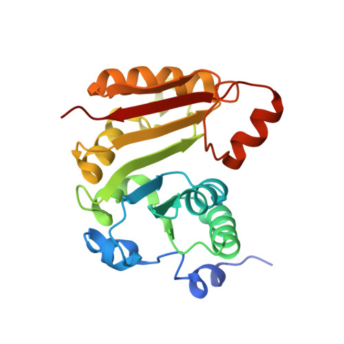
Structural model of the M7G46 Methyltransferase TrmB in complex with tRNA.
Blersch, K.F., Burchert, J.P., August, S.C., Welp, L., Neumann, P., Koster, S., Urlaub, H., Ficner, R.(2021) RNA Biol 18: 2466-2479
- PubMed: 34006170 Search on PubMedSearch on PubMed Central
- DOI: https://doi.org/10.1080/15476286.2021.1925477
- Primary Citation Related Structures:
7NYB, 7NZI, 7NZJ - PubMed Abstract:
TrmB belongs to the class I S-adenosylmethionine (SAM)-dependent methyltransferases (MTases) and introduces a methyl group to guanine at position 7 (m 7 G) in tRNA. In tRNAs m 7 G is most frequently found at position 46 in the variable loop and forms a tertiary base pair with C13 and U22, introducing a positive charge at G46. The TrmB/Trm8 enzyme family is structurally diverse, as TrmB proteins exist in a monomeric, homodimeric, and heterodimeric form. So far, the exact enzymatic mechanism, as well as the tRNA-TrmB crystal structure is not known. Here we present the first crystal structures of B. subtilis TrmB in complex with SAM and SAH. The crystal structures of TrmB apo and in complex with SAM and SAH have been determined by X-ray crystallography to 1.9 Å (apo), 2.5 Å (SAM), and 3.1 Å (SAH). The obtained crystal structures revealed Tyr193 to be important during SAM binding and MTase activity. Applying fluorescence polarization, the dissociation constant K d of TrmB and tRNA Phe was determined to be 0.12 µM ± 0.002 µM. Luminescence-based methyltransferase activity assays revealed cooperative effects during TrmB catalysis with half-of-the-site reactivity at physiological SAM concentrations. Structural data retrieved from small-angle x-ray scattering (SAXS), mass-spectrometry of cross-linked complexes, and molecular docking experiments led to the determination of the TrmB-tRNA Phe complex structure.
- Department of Molecular Structural Biology, Institute of Microbiology and Genetics, GZMB, Georg August University Göttingen, Göttingen, Germany.
Organizational Affiliation: