7R63 | pdb_00007r63

Nb82, a nanobody against voltage gated sodium channels Nav1.4 and Nav1.5


Experimental Data Snapshot

  • Method: X-RAY DIFFRACTION
  • Resolution: 2.00 Å
  • R-Value Free: 
    0.244 (Depositor), 0.250 (DCC) 
  • R-Value Work: 
    0.184 (Depositor) 
  • R-Value Observed: 
    0.187 (Depositor) 

Starting Model: experimental
View more details

wwPDB Validation 3D Report Full Report

Validation slider image for 7R63

This is version 1.2 of the entry. See complete history. 

Literature

Development of high-affinity nanobodies specific for Na V 1.4 and Na V 1.5 voltage-gated sodium channel isoforms.

Srinivasan, L., Alzogaray, V., Selvakumar, D., Nathan, S., Yoder, J.B., Wright, K.M., Klinke, S., Nwafor, J.N., Labanda, M.S., Goldbaum, F.A., Schon, A., Freire, E., Tomaselli, G.F., Amzel, L.M., Ben-Johny, M., Gabelli, S.B.

(2022) J Biol Chem 298: 101763-101763

  • DOI: https://doi.org/10.1016/j.jbc.2022.101763
  • Primary Citation Related Structures: 
    7R63

  • PubMed Abstract: 

    Voltage-gated sodium channels, Na V s, are responsible for the rapid rise of action potentials in excitable tissues. Na V channel mutations have been implicated in several human genetic diseases, such as hypokalemic periodic paralysis, myotonia, and long-QT and Brugada syndromes. Here, we generated high-affinity anti-Na V nanobodies (Nbs), Nb17 and Nb82, that recognize the Na V 1.4 (skeletal muscle) and Na V 1.5 (cardiac muscle) channel isoforms. These Nbs were raised in llama (Lama glama) and selected from a phage display library for high affinity to the C-terminal (CT) region of Na V 1.4. The Nbs were expressed in Escherichia coli, purified, and biophysically characterized. Development of high-affinity Nbs specifically targeting a given human Na V isoform has been challenging because they usually show undesired crossreactivity for different Na V isoforms. Our results show, however, that Nb17 and Nb82 recognize the CTNa V 1.4 or CTNa V 1.5 over other CTNav isoforms. Kinetic experiments by biolayer interferometry determined that Nb17 and Nb82 bind to the CTNa V 1.4 and CTNa V 1.5 with high affinity (K D ∼ 40-60 nM). In addition, as proof of concept, we show that Nb82 could detect Na V 1.4 and Na V 1.5 channels in mammalian cells and tissues by Western blot. Furthermore, human embryonic kidney cells expressing holo Na V 1.5 channels demonstrated a robust FRET-binding efficiency for Nb17 and Nb82. Our work lays the foundation for developing Nbs as anti-Na V reagents to capture Na V s from cell lysates and as molecular visualization agents for Na V s.


  • Organizational Affiliation: 
    • Department of Biophysics and Biophysical Chemistry, The Johns Hopkins School of Medicine, Baltimore, Maryland, USA.

Macromolecule Content 

  • Total Structure Weight: 62.14 kDa 
  • Atom Count: 4,363 
  • Modeled Residue Count: 513 
  • Deposited Residue Count: 548 
  • Unique protein chains: 1

Macromolecules

Find similar proteins by:|  3D Structure
Entity ID: 1
MoleculeChains  Sequence LengthOrganismDetailsImage
Nanobody Nb82
A, B, C, D
137Lama glamaMutation(s): 0 
Gene Names: VHH
Entity Groups
Sequence Clusters30% Identity50% Identity70% Identity90% Identity95% Identity100% Identity
Sequence Annotations
Expand
Reference Sequence

Small Molecules

Ligands 4 Unique
IDChains Name / Formula / InChI Key2D Diagram3D Interactions
PG0

Query on PG0


Download:Ideal Coordinates CCD File
H [auth A],
M [auth B],
T [auth D]
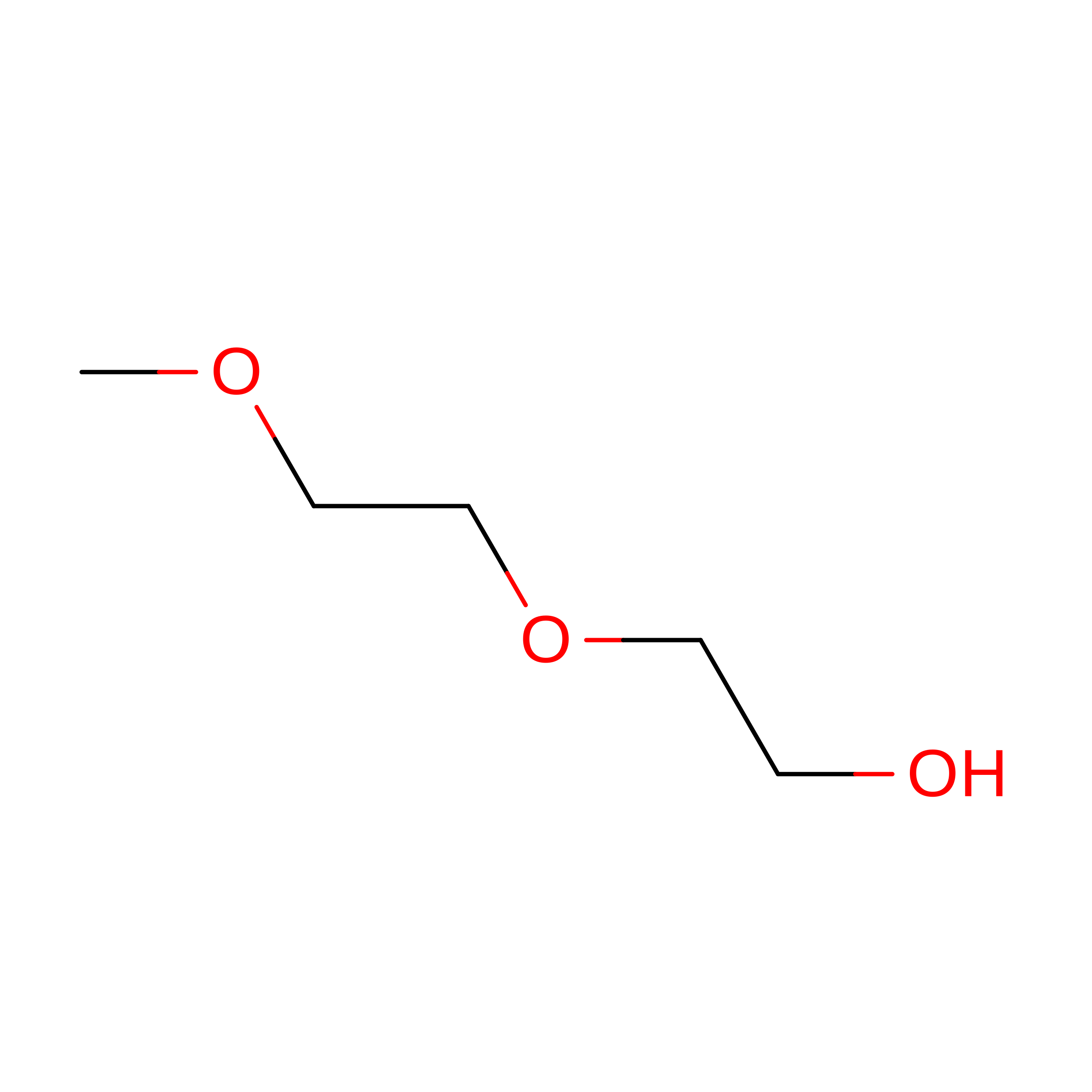
2-(2-METHOXYETHOXY)ETHANOL
C5 H12 O3
SBASXUCJHJRPEV-UHFFFAOYSA-N
SO4

Query on SO4


Download:Ideal Coordinates CCD File
E [auth A]
F [auth A]
G [auth A]
K [auth B]
L [auth B]
E [auth A],
F [auth A],
G [auth A],
K [auth B],
L [auth B],
O [auth C],
R [auth D],
S [auth D]
SULFATE ION
O4 S
QAOWNCQODCNURD-UHFFFAOYSA-L
CL

Query on CL


Download:Ideal Coordinates CCD File
P [auth C]CHLORIDE ION
Cl
VEXZGXHMUGYJMC-UHFFFAOYSA-M
NA

Query on NA


Download:Ideal Coordinates CCD File
I [auth B],
J [auth B],
N [auth C],
Q [auth D]
SODIUM ION
Na
FKNQFGJONOIPTF-UHFFFAOYSA-N

Experimental Data & Validation

Experimental Data

  • Method: X-RAY DIFFRACTION
  • Resolution: 2.00 Å
  • R-Value Free:  0.244 (Depositor), 0.250 (DCC) 
  • R-Value Work:  0.184 (Depositor) 
  • R-Value Observed: 0.187 (Depositor) 
Space Group: C 2 2 21
Unit Cell:
Length ( Å )Angle ( ˚ )
a = 77.745α = 90
b = 82.474β = 90
c = 170.7γ = 90
Software Package:
Software NamePurpose
Aimlessdata scaling
REFMACrefinement
PDB_EXTRACTdata extraction
XDSdata reduction
PHASERphasing

Structure Validation

View Full Validation Report


Entry History 

& Funding Information

Deposition Data


Funding OrganizationLocationGrant Number
National Institutes of Health/National Heart, Lung, and Blood Institute (NIH/NHLBI)United StatesR01 HLI28743-01A1

Revision History  (Full details and data files)

  • Version 1.0: 2022-05-04
    Type: Initial release
  • Version 1.1: 2023-10-18
    Changes: Data collection, Refinement description
  • Version 1.2: 2024-10-30
    Changes: Structure summary