The beta-latch structural element of the SufS cysteine desulfurase mediates active site accessibility and SufE transpersulfurase positioning.
Gogar, R.K., Carroll, F., Conte, J.V., Nasef, M., Dunkle, J.A., Frantom, P.A.(2023) J Biological Chem 299: 102966-102966
- PubMed: 36736428 Search on PubMedSearch on PubMed Central
- DOI: https://doi.org/10.1016/j.jbc.2023.102966
- Primary Citation Related Structures:
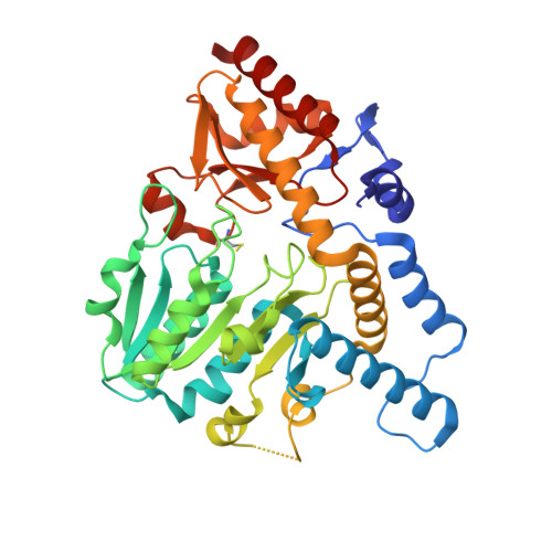
7RUJ, 7RW3 - PubMed Abstract:
Under oxidative stress and iron starvation conditions, Escherichia coli uses the Suf pathway to assemble iron-sulfur clusters. The Suf pathway mobilizes sulfur via SufS, a type II cysteine desulfurase. SufS is a pyridoxal-5'-phosphate-dependent enzyme that uses cysteine to generate alanine and an active-site persulfide (C 364 -S-S - ). The SufS persulfide is protected from external oxidants/reductants and requires the transpersulfurase, SufE, to accept the persulfide to complete the SufS catalytic cycle. Recent reports on SufS identified a conserved "β-latch" structural element that includes the α 6 helix, a glycine-rich loop, a β-hairpin, and a cis-proline residue. To identify a functional role for the β-latch, we used site-directed mutagenesis to obtain the N99D and N99A SufS variants. N99 is a conserved residue that connects the α 6 helix to the backbone of the glycine-rich loop via hydrogen bonds. Our x-ray crystal structures for N99A and N99D SufS show a distorted beta-hairpin and glycine-rich loop, respectively, along with changes in the dimer geometry. The structural disruption of the N99 variants allowed the external reductant TCEP to react with the active-site C364-persulfide intermediate to complete the SufS catalytic cycle in the absence of SufE. The substitutions also appear to disrupt formation of a high-affinity, close approach SufS-SufE complex as measured with fluorescence polarization. Collectively, these findings demonstrate that the β-latch does not affect the chemistry of persulfide formation but does protect it from undesired reductants. The data also indicate the β-latch plays an unexpected role in forming a close approach SufS-SufE complex to promote persulfide transfer.
- Department of Chemistry & Biochemistry, The University of Alabama, Tuscaloosa, Alabama, USA.
Organizational Affiliation: