7S8G

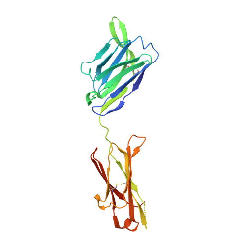
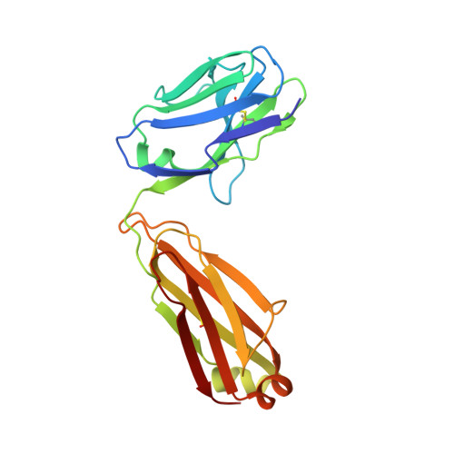
Structure of anti-LASV Fab 25.10C with FNQI mutation


Experimental Data Snapshot

  • Method: X-RAY DIFFRACTION
  • Resolution: 2.57 Å
  • R-Value Free: 0.263 
  • R-Value Work: 0.220 
  • R-Value Observed: 0.225 

Starting Model: experimental
View more details

wwPDB Validation   3D Report Full Report


This is version 1.3 of the entry. See complete history


Literature

Delineating the mechanism of anti-Lassa virus GPC-A neutralizing antibodies.

Enriquez, A.S.Buck, T.K.Li, H.Norris, M.J.Moon-Walker, A.Zandonatti, M.A.Harkins, S.S.Robinson, J.E.Branco, L.M.Garry, R.F.Saphire, E.O.Hastie, K.M.

(2022) Cell Rep 39: 110841-110841

  • DOI: https://doi.org/10.1016/j.celrep.2022.110841
  • Primary Citation of Related Structures:  
    7S8G, 7S8H, 7TYV

  • PubMed Abstract: 

    Lassa virus (LASV) is the etiologic agent of Lassa Fever, a hemorrhagic disease that is endemic to West Africa. During LASV infection, LASV glycoprotein (GP) engages with multiple host receptors for cell entry. Neutralizing antibodies against GP are rare and principally target quaternary epitopes displayed only on the metastable, pre-fusion conformation of GP. Currently, the structural features of the neutralizing GPC-A antibody competition group are understudied. Structures of two GPC-A antibodies presented here demonstrate that they bind the side of the pre-fusion GP trimer, bridging the GP1 and GP2 subunits. Complementary biochemical analyses indicate that antibody 25.10C, which is broadly specific, neutralizes by inhibiting binding of the endosomal receptor LAMP1 and also by blocking membrane fusion. The other GPC-A antibody, 36.1F, which is lineage-specific, prevents LAMP1 association only. These data illuminate a site of vulnerability on LASV GP and will guide efforts to elicit broadly reactive therapeutics and vaccines.


  • Organizational Affiliation

    La Jolla Institute for Immunology, La Jolla, CA 92037, USA.


Macromolecules
Find similar proteins by:  (by identity cutoff)  |  3D Structure
Entity ID: 1
MoleculeChains Sequence LengthOrganismDetailsImage
25.10C-FNQI Fab Heavy ChainA [auth H],
C [auth h]
227Homo sapiensMutation(s): 0 
Entity Groups  
Sequence Clusters30% Identity50% Identity70% Identity90% Identity95% Identity100% Identity
Sequence Annotations
Expand
  • Reference Sequence
Find similar proteins by:  (by identity cutoff)  |  3D Structure
Entity ID: 2
MoleculeChains Sequence LengthOrganismDetailsImage
25.10C-FNQI Fab Light ChainB [auth L],
D [auth l]
210Homo sapiensMutation(s): 0 
Entity Groups  
Sequence Clusters30% Identity50% Identity70% Identity90% Identity95% Identity100% Identity
Sequence Annotations
Expand
  • Reference Sequence
Experimental Data & Validation

Experimental Data

  • Method: X-RAY DIFFRACTION
  • Resolution: 2.57 Å
  • R-Value Free: 0.263 
  • R-Value Work: 0.220 
  • R-Value Observed: 0.225 
  • Space Group: P 1 21 1
Unit Cell:
Length ( Å )Angle ( ˚ )
a = 71.6α = 90
b = 72.718β = 113.267
c = 90.014γ = 90
Software Package:
Software NamePurpose
PHENIXrefinement
XDSdata reduction
Aimlessdata scaling
PHENIXphasing

Structure Validation

View Full Validation Report


Entry History & Funding Information

Deposition Data


Funding OrganizationLocationGrant Number
National Institutes of Health/National Institute Of Allergy and Infectious Diseases (NIH/NIAID)United StatesU19A142790

Revision History  (Full details and data files)

  • Version 1.0: 2022-06-01
    Type: Initial release
  • Version 1.1: 2022-06-15
    Changes: Database references
  • Version 1.2: 2023-10-18
    Changes: Data collection, Refinement description
  • Version 1.3: 2024-11-20
    Changes: Structure summary