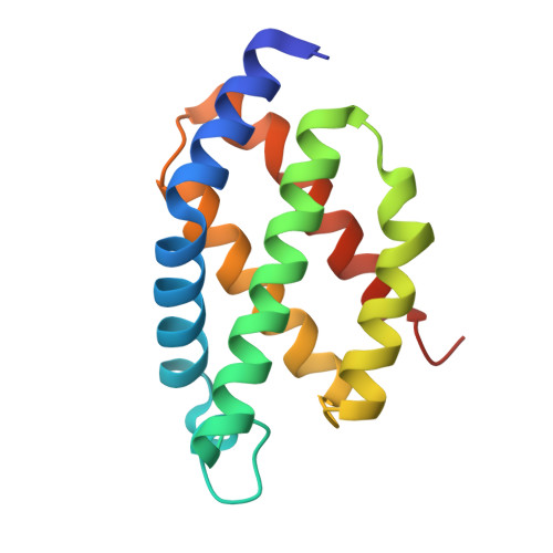
7UQA | pdb_00007uqa

Crystal structure of the small Ultra-Red Fluorescent Protein (smURFP)


Experimental Data Snapshot

  • Method: X-RAY DIFFRACTION
  • Resolution: 2.80 Å
  • R-Value Free: 
    0.263 (Depositor), 0.268 (DCC) 
  • R-Value Work: 
    0.239 (Depositor), 0.242 (DCC) 
  • R-Value Observed: 
    0.241 (Depositor) 

Starting Model: experimental
View more details

wwPDB Validation 3D Report Full Report

Validation slider image for 7UQA

This is version 1.1 of the entry. See complete history

Literature

Structural and photophysical characterization of the small ultra-red fluorescent protein.

Maiti, A.Buffalo, C.Z.Saurabh, S.Montecinos-Franjola, F.Hachey, J.S.Conlon, W.J.Tran, G.N.Hassan, B.Walters, K.J.Drobizhev, M.Moerner, W.E.Ghosh, P.Matsuo, H.Tsien, R.Y.Lin, J.Y.Rodriguez, E.A.

(2023) Nat Commun 14: 4155-4155

  • DOI: https://doi.org/10.1038/s41467-023-39776-9
  • Primary Citation Related Structures: 
    7UQA

  • PubMed Abstract: 

    The small Ultra-Red Fluorescent Protein (smURFP) represents a new class of fluorescent protein with exceptional photostability and brightness derived from allophycocyanin in a previous directed evolution. Here, we report the smURFP crystal structure to better understand properties and enable further engineering of improved variants. We compare this structure to the structures of allophycocyanin and smURFP mutants to identify the structural origins of the molecular brightness. We then use a structure-guided approach to develop monomeric smURFP variants that fluoresce with phycocyanobilin but not biliverdin. Furthermore, we measure smURFP photophysical properties necessary for advanced imaging modalities, such as those relevant for two-photon, fluorescence lifetime, and single-molecule imaging. We observe that smURFP has the largest two-photon cross-section measured for a fluorescent protein, and that it produces more photons than organic dyes. Altogether, this study expands our understanding of the smURFP, which will inform future engineering toward optimal FPs compatible with whole organism studies.


  • Organizational Affiliation
    • Cancer Innovation Laboratory, Frederick National Laboratory for Cancer Research, Frederick, MD, 21702, USA.

Macromolecule Content 

  • Total Structure Weight: 96.6 kDa 
  • Atom Count: 6,170 
  • Modeled Residue Count: 793 
  • Deposited Residue Count: 840 
  • Unique protein chains: 1

Macromolecules

Find similar proteins by:|  3D Structure
Entity ID: 1
MoleculeChains  Sequence LengthOrganismDetailsImage
small Ultra-Red Fluorescent Protein (smURFP)
A, B, C, D, E
A, B, C, D, E, F
140synthetic constructMutation(s): 0 

Small Molecules

Ligands 2 Unique
IDChains Name / Formula / InChI Key2D Diagram3D Interactions
CL

Query on CL


Download:Ideal Coordinates CCD File
G [auth A]
H [auth A]
I [auth C]
J [auth C]
K [auth C]
G [auth A],
H [auth A],
I [auth C],
J [auth C],
K [auth C],
M [auth D],
N [auth E],
O [auth E],
P [auth E],
Q [auth F],
R [auth F],
S [auth F],
T [auth F],
U [auth F]
CHLORIDE ION
Cl
VEXZGXHMUGYJMC-UHFFFAOYSA-M
NA

Query on NA


Download:Ideal Coordinates CCD File
L [auth C]SODIUM ION
Na
FKNQFGJONOIPTF-UHFFFAOYSA-N

Experimental Data & Validation

Experimental Data

  • Method: X-RAY DIFFRACTION
  • Resolution: 2.80 Å
  • R-Value Free:  0.263 (Depositor), 0.268 (DCC) 
  • R-Value Work:  0.239 (Depositor), 0.242 (DCC) 
  • R-Value Observed: 0.241 (Depositor) 
Space Group: C 2 2 21
Unit Cell:
Length ( Å )Angle ( ˚ )
a = 81.087α = 90
b = 141.802β = 90
c = 171.942γ = 90
Software Package:
Software NamePurpose
HKL-2000data scaling
PHENIXrefinement
PDB_EXTRACTdata extraction
HKL-2000data reduction
PHASERphasing

Structure Validation

View Full Validation Report


Entry History 

& Funding Information

Deposition Data


Funding OrganizationLocationGrant Number
National Institutes of Health/National Institute of Neurological Disorders and Stroke (NIH/NINDS)United StatesU01NS090590
National Institutes of Health/National Institute of General Medical Sciences (NIH/NIGMS)United StatesR01GM086197
National Institutes of Health/National Cancer Institute (NIH/NCI)United StatesHHSN26120080001E

Revision History  (Full details and data files)

  • Version 1.0: 2023-07-19
    Type: Initial release
  • Version 1.1: 2023-10-25
    Changes: Data collection, Refinement description