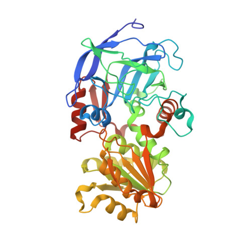
7UTW

Cd-substituted Horse Liver Alcohol Dehydrogenase in Complex with NADH and N-Cyclohexylformamide


Experimental Data Snapshot

  • Method: X-RAY DIFFRACTION
  • Resolution: 1.33 Å
  • R-Value Free: 0.165 
  • R-Value Work: 0.138 
  • R-Value Observed: 0.139 

Starting Model: experimental
View more details

wwPDB Validation   3D Report Full Report


Ligand Structure Quality Assessment 


This is version 1.3 of the entry. See complete history


Literature

Enhanced active-site electric field accelerates enzyme catalysis.

Zheng, C.Ji, Z.Mathews, I.I.Boxer, S.G.

(2023) Nat Chem 15: 1715-1721

  • DOI: https://doi.org/10.1038/s41557-023-01287-x
  • Primary Citation of Related Structures:  
    7U9N, 7UQ9, 7UTW, 8EIW, 8EIX, 8EIY

  • PubMed Abstract: 

    The design and improvement of enzymes based on physical principles remain challenging. Here we demonstrate that the principle of electrostatic catalysis can be leveraged to substantially improve a natural enzyme's activity. We enhanced the active-site electric field in horse liver alcohol dehydrogenase by replacing the serine hydrogen-bond donor with threonine and replacing the catalytic Zn 2+ with Co 2+ . Based on the electric field enhancement, we make a quantitative prediction of rate acceleration-50-fold faster than the wild-type enzyme-which was in close agreement with experimental measurements. The effects of the hydrogen bonding and metal coordination, two distinct chemical forces, are described by a unified physical quantity-electric field, which is quantitative, and shown here to be additive and predictive. These results suggest a new design paradigm for both biological and non-biological catalysts.


  • Organizational Affiliation

    Department of Chemistry, Stanford University, Stanford, CA, USA.


Macromolecules
Find similar proteins by:  (by identity cutoff)  |  3D Structure
Entity ID: 1
MoleculeChains Sequence LengthOrganismDetailsImage
Alcohol dehydrogenase E chain
A, B
377Equus caballusMutation(s): 0 
EC: 1.1.1.1
UniProt
Find proteins for P00327 (Equus caballus)
Explore P00327 
Go to UniProtKB:  P00327
Entity Groups  
Sequence Clusters30% Identity50% Identity70% Identity90% Identity95% Identity100% Identity
UniProt GroupP00327
Sequence Annotations
Expand
  • Reference Sequence
Small Molecules
Ligands 4 Unique
IDChains Name / Formula / InChI Key2D Diagram3D Interactions
NAI
Query on NAI

Download Ideal Coordinates CCD File 
E [auth A],
I [auth B]
1,4-DIHYDRONICOTINAMIDE ADENINE DINUCLEOTIDE
C21 H29 N7 O14 P2
BOPGDPNILDQYTO-NNYOXOHSSA-N
CXF (Subject of Investigation/LOI)
Query on CXF

Download Ideal Coordinates CCD File 
F [auth A],
J [auth B]
CYCLOHEXYLFORMAMIDE
C7 H13 N O
SWGXDLRCJNEEGZ-UHFFFAOYSA-N
CD
Query on CD

Download Ideal Coordinates CCD File 
C [auth A],
G [auth B]
CADMIUM ION
Cd
WLZRMCYVCSSEQC-UHFFFAOYSA-N
ZN
Query on ZN

Download Ideal Coordinates CCD File 
D [auth A],
H [auth B]
ZINC ION
Zn
PTFCDOFLOPIGGS-UHFFFAOYSA-N
Experimental Data & Validation

Experimental Data

  • Method: X-RAY DIFFRACTION
  • Resolution: 1.33 Å
  • R-Value Free: 0.165 
  • R-Value Work: 0.138 
  • R-Value Observed: 0.139 
  • Space Group: P 1
Unit Cell:
Length ( Å )Angle ( ˚ )
a = 44.206α = 92.39
b = 50.807β = 102.93
c = 92.903γ = 109.33
Software Package:
Software NamePurpose
Aimlessdata scaling
PHENIXrefinement
PDB_EXTRACTdata extraction
XDSdata reduction
PHENIXphasing

Structure Validation

View Full Validation Report


Ligand Structure Quality Assessment 


Entry History & Funding Information

Deposition Data


Funding OrganizationLocationGrant Number
National Institutes of Health/National Institute of General Medical Sciences (NIH/NIGMS)United StatesGM118044

Revision History  (Full details and data files)

  • Version 1.0: 2023-02-01
    Type: Initial release
  • Version 1.1: 2023-10-25
    Changes: Data collection, Refinement description
  • Version 1.2: 2023-12-06
    Changes: Database references
  • Version 1.3: 2023-12-13
    Changes: Database references