7VE3

Structure of the complex of sheep lactoperoxidase with hypoiodite at 2.70 A resolution


Experimental Data Snapshot

  • Method: X-RAY DIFFRACTION
  • Resolution: 2.70 Å
  • R-Value Free: 0.277 
  • R-Value Work: 0.195 

wwPDB Validation   3D Report Full Report


Ligand Structure Quality Assessment 


This is version 1.3 of the entry. See complete history


Literature

Structural evidence of the oxidation of iodide ion into hyper-reactive hypoiodite ion by mammalian heme lactoperoxidase.

Singh, P.K.Ahmad, N.Yamini, S.Singh, R.P.Singh, A.K.Sharma, P.Smith, M.L.Sharma, S.Singh, T.P.

(2022) Protein Sci 31: 384-395

  • DOI: https://doi.org/10.1002/pro.4230
  • Primary Citation of Related Structures:  
    7VE3

  • PubMed Abstract: 

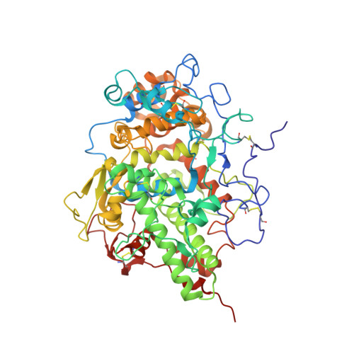
    Lactoperoxidase (1.11.1.7, LPO) is a mammalian heme peroxidase found in the extracellular fluids of mammals including plasma, saliva, airway epithelial lining fluids, nasal lining fluid, milk, tears, gastric juices, and intestinal mucosa. To perform its innate immune action against invading microbes, LPO utilizes hydrogen peroxide (H 2 O 2 ) to convert thiocyanate (SCN - ) and iodide (I - ) ions into the oxidizing compounds hypothiocyanite (OSCN - ) and hypoiodite (IO - ). Previously determined structures of the complexes of LPO with SCN - , OSCN - , and I - show that SCN - and I - occupy appropriate positions in the distal heme cavity as substrates while OSCN - binds in the distal heme cavity as a product inhibitor. We report here the structure of the complex of LPO with IO - as the first structural evidence of the conversion of iodide into hypoiodite by LPO. To obtain this complex, a solution of LPO was first incubated with H 2 O 2 , then mixed with ammonium iodide solution and the complex crystallized by the addition of PEG-3350, 20% (wt/vol). These crystals were used for X-ray intensity data collection and structure analysis. The structure determination revealed the presence of four hypoiodite ions in the substrate binding channel of LPO. In addition to these, six other hypoiodite ions were observed at different exterior sites. We surmise that the presence of hypoiodite ions in the distal heme cavity blocks the substrate binding site and inhibits catalysis. This was confirmed by activity experiments with the colorimetric substrate, ABTS (2,2'-azino-bis(3-ethylbenzthiazoline-sulfonic acid)), in the presence of hypoiodite and iodide ions.


  • Organizational Affiliation

    Department of Biophysics, All India Institute of Medical Sciences, New Delhi, India.


Macromolecules
Find similar proteins by:  (by identity cutoff)  |  3D Structure
Entity ID: 1
MoleculeChains Sequence LengthOrganismDetailsImage
Lactoperoxidase595Ovis ariesMutation(s): 0 
EC: 1.11.1.7
Entity Groups  
Sequence Clusters30% Identity50% Identity70% Identity90% Identity95% Identity100% Identity
Sequence Annotations
Expand
  • Reference Sequence
Small Molecules
Ligands 6 Unique
IDChains Name / Formula / InChI Key2D Diagram3D Interactions
HEM (Subject of Investigation/LOI)
Query on HEM

Download Ideal Coordinates CCD File 
B [auth A]PROTOPORPHYRIN IX CONTAINING FE
C34 H32 Fe N4 O4
KABFMIBPWCXCRK-RGGAHWMASA-L
NAG (Subject of Investigation/LOI)
Query on NAG

Download Ideal Coordinates CCD File 
D [auth A],
E [auth A]
2-acetamido-2-deoxy-beta-D-glucopyranose
C8 H15 N O6
OVRNDRQMDRJTHS-FMDGEEDCSA-N
61D (Subject of Investigation/LOI)
Query on 61D

Download Ideal Coordinates CCD File 
M [auth A]
N [auth A]
O [auth A]
P [auth A]
Q [auth A]
M [auth A],
N [auth A],
O [auth A],
P [auth A],
Q [auth A],
R [auth A],
S [auth A],
T [auth A],
U [auth A],
V [auth A],
W [auth A]
hypoiodous acid
H I O
GEOVEUCEIQCBKH-UHFFFAOYSA-N
IOD
Query on IOD

Download Ideal Coordinates CCD File 
F [auth A]
G [auth A]
H [auth A]
I [auth A]
J [auth A]
F [auth A],
G [auth A],
H [auth A],
I [auth A],
J [auth A],
K [auth A]
IODIDE ION
I
XMBWDFGMSWQBCA-UHFFFAOYSA-M
SO4
Query on SO4

Download Ideal Coordinates CCD File 
L [auth A]SULFATE ION
O4 S
QAOWNCQODCNURD-UHFFFAOYSA-L
CA
Query on CA

Download Ideal Coordinates CCD File 
C [auth A]CALCIUM ION
Ca
BHPQYMZQTOCNFJ-UHFFFAOYSA-N
Experimental Data & Validation

Experimental Data

  • Method: X-RAY DIFFRACTION
  • Resolution: 2.70 Å
  • R-Value Free: 0.277 
  • R-Value Work: 0.195 
  • Space Group: P 1 21 1
Unit Cell:
Length ( Å )Angle ( ˚ )
a = 53.942α = 90
b = 80.179β = 102.86
c = 76.122γ = 90
Software Package:
Software NamePurpose
REFMACrefinement
DENZOdata reduction
SCALEPACKdata scaling
AMoREphasing

Structure Validation

View Full Validation Report


Ligand Structure Quality Assessment 


Entry History & Funding Information

Deposition Data


Funding OrganizationLocationGrant Number
Not funded--

Revision History  (Full details and data files)

  • Version 1.0: 2021-09-22
    Type: Initial release
  • Version 1.1: 2021-12-01
    Changes: Database references
  • Version 1.2: 2022-02-23
    Changes: Database references
  • Version 1.3: 2023-11-29
    Changes: Data collection, Derived calculations, Refinement description