7VNQ

Structure of human KCNQ4-ML213 complex in nanodisc


Experimental Data Snapshot

  • Method: ELECTRON MICROSCOPY
  • Resolution: 2.96 Å
  • Aggregation State: PARTICLE 
  • Reconstruction Method: SINGLE PARTICLE 

wwPDB Validation   3D Report Full Report


This is version 1.1 of the entry. See complete history


Literature

Structural insights into the lipid and ligand regulation of a human neuronal KCNQ channel.

Zheng, Y.Liu, H.Chen, Y.Dong, S.Wang, F.Wang, S.Li, G.L.Shu, Y.Xu, F.

(2022) Neuron 110: 237

  • DOI: https://doi.org/10.1016/j.neuron.2021.10.029
  • Primary Citation of Related Structures:  
    7VNP, 7VNQ, 7VNR

  • PubMed Abstract: 

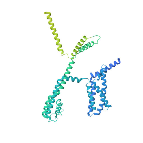
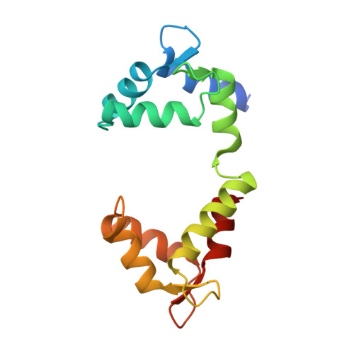
    The KCNQ family (KCNQ1-KCNQ5) of voltage-gated potassium channels plays critical roles in many physiological and pathological processes. It is known that the channel opening of all KCNQs relies on the signaling lipid molecule phosphatidylinositol 4,5-bisphosphate (PIP2). However, the molecular mechanism of PIP2 in modulating the opening of the four neuronal KCNQ channels (KCNQ2-KCNQ5), which are essential for regulating neuronal excitability, remains largely elusive. Here, we report the cryoelectron microscopy (cryo-EM) structures of human KCNQ4 determined in complex with the activator ML213 in the absence or presence of PIP2. Two PIP2 molecules are identified in the open-state structure of KCNQ4, which act as a bridge to couple the voltage-sensing domain (VSD) and pore domain (PD) of KCNQ4 leading to the channel opening. Our findings reveal the binding sites and activation mechanisms of ML213 and PIP2 for neuronal KCNQ channels, providing a framework for therapeutic intervention targeting on these important channels.


  • Organizational Affiliation

    iHuman Institute, ShanghaiTech University, Shanghai, China; School of Life Science and Technology, ShanghaiTech University, Shanghai, China; Center for Excellence in Molecular Cell Science, Shanghai Institutes for Biological Sciences, Chinese Academy of Sciences, Shanghai, China; University of Chinese Academy of Sciences, Beijing, China.


Macromolecules
Find similar proteins by:  (by identity cutoff)  |  3D Structure
Entity ID: 1
MoleculeChains Sequence LengthOrganismDetailsImage
Potassium voltage-gated channel subfamily KQT member 4,Maltodextrin-binding protein
A, C, E, G
1,049Homo sapiensEscherichia coli BL21(DE3)
This entity is chimeric
Mutation(s): 0 
Gene Names: KCNQ4ECBD_4002
Membrane Entity: Yes 
UniProt & NIH Common Fund Data Resources
Find proteins for P56696 (Homo sapiens)
Explore P56696 
Go to UniProtKB:  P56696
PHAROS:  P56696
GTEx:  ENSG00000117013 
Entity Groups  
Sequence Clusters30% Identity50% Identity70% Identity90% Identity95% Identity100% Identity
UniProt GroupP56696
Sequence Annotations
Expand
  • Reference Sequence
Find similar proteins by:  (by identity cutoff)  |  3D Structure
Entity ID: 2
MoleculeChains Sequence LengthOrganismDetailsImage
Calmodulin-3
B, D, F, H
149Homo sapiensMutation(s): 0 
Gene Names: CALM3CALML2CAM3CAMCCAMIII
UniProt & NIH Common Fund Data Resources
Find proteins for P0DP25 (Homo sapiens)
Explore P0DP25 
Go to UniProtKB:  P0DP25
PHAROS:  P0DP25
GTEx:  ENSG00000160014 
Entity Groups  
Sequence Clusters30% Identity50% Identity70% Identity90% Identity95% Identity100% Identity
UniProt GroupP0DP25
Sequence Annotations
Expand
  • Reference Sequence
Experimental Data & Validation

Experimental Data

  • Method: ELECTRON MICROSCOPY
  • Resolution: 2.96 Å
  • Aggregation State: PARTICLE 
  • Reconstruction Method: SINGLE PARTICLE 

Structure Validation

View Full Validation Report


Entry History & Funding Information

Deposition Data


Funding OrganizationLocationGrant Number
Ministry of Science and Technology (MoST, China)China2018YFA0507000

Revision History  (Full details and data files)

  • Version 1.0: 2021-12-01
    Type: Initial release
  • Version 1.1: 2022-02-16
    Changes: Database references