Structural and biochemical insights into purine-based drug molecules in hBRD2 delineate a unique binding mode opening new vistas in the design of inhibitors of the BET family.
Arole, A.H., Deshmukh, P., Sridhar, A., Mathur, S., Mahalingaswamy, M., Subramanya, H., Dalavaikodihalli Nanjaiah, N., Padmanabhan, B.(2023) Acta Crystallogr D Struct Biol 79: 758-774
- PubMed: 37432115 Search on PubMed
- DOI: https://doi.org/10.1107/S2059798323005211
- Primary Citation Related Structures:
7VRH, 7VRI, 7VRK, 7VRM, 7VRO, 7VRQ, 7VRZ, 7VS0, 7VS1, 7VSF - PubMed Abstract:
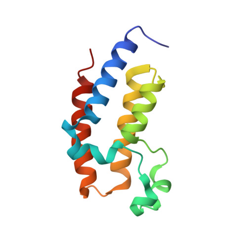
The bromodomain and extra-terminal (BET) family proteins, which are involved in chromatin function, have been shown to be promising drug targets in several pathological conditions, including cancer and inflammation. There is considerable interest in the development of BET inhibitors with novel scaffolds to modulate the epigenesis of such diseases. Here, high-resolution crystal structures of the purine class of FDA-approved drugs (theophylline, doxophylline and acyclovir) and non-FDA-approved compounds (3-methyl-7-propylxanthine and theobromine) complexed with hBRD2 bromodomains BD1 and BD2 are reported. Remarkably, a new binding site is exhibited by stacking the compounds against the WPF shelf of BD1 and BD2. This serendipitous binding, in addition to the known acetyl-lysine binding site, sufficiently anchors the ligands in the solvent-exposed region. In addition, slight variations in the lipophilicity of these molecules significantly affected the in vitro binding affinity and selectivity towards BD1 compared with BD2. This idiosyncratic binding provides a new structural framework to link these sites for the development of next-generation inhibitors of the BET family.
- Department of Biophysics, National Institute of Mental Health and Neurosciences (NIMHANS), Hosur Main Road, Bengaluru 560029, India.
Organizational Affiliation: