7WFW

Apo human Nav1.8


Experimental Data Snapshot

  • Method: ELECTRON MICROSCOPY
  • Resolution: 3.10 Å
  • Aggregation State: PARTICLE 
  • Reconstruction Method: SINGLE PARTICLE 

wwPDB Validation   3D Report Full Report


This is version 1.1 of the entry. See complete history


Literature

Structural basis for high-voltage activation and subtype-specific inhibition of human Na v 1.8.

Huang, X.Jin, X.Huang, G.Huang, J.Wu, T.Li, Z.Chen, J.Kong, F.Pan, X.Yan, N.

(2022) Proc Natl Acad Sci U S A 119: e2208211119-e2208211119

  • DOI: https://doi.org/10.1073/pnas.2208211119
  • Primary Citation of Related Structures:  
    7WFW

  • PubMed Abstract: 

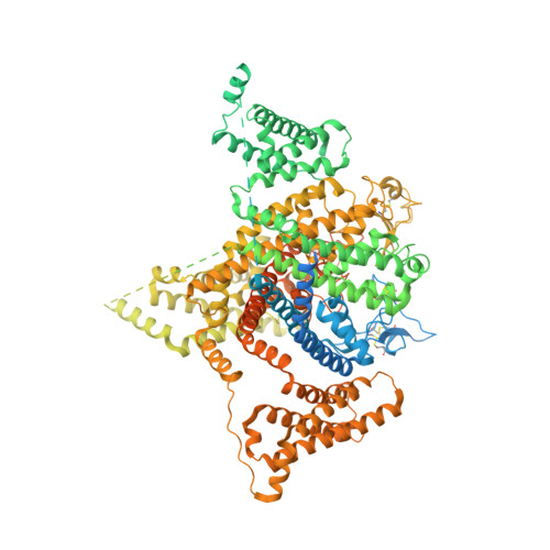
    The dorsal root ganglia-localized voltage-gated sodium (Na v ) channel Na v 1.8 represents a promising target for developing next-generation analgesics. A prominent characteristic of Na v 1.8 is the requirement of more depolarized membrane potential for activation. Here we present the cryogenic electron microscopy structures of human Na v 1.8 alone and bound to a selective pore blocker, A-803467, at overall resolutions of 2.7 to 3.2 Å. The first voltage-sensing domain (VSD I ) displays three different conformations. Structure-guided mutagenesis identified the extracellular interface between VSD I and the pore domain (PD) to be a determinant for the high-voltage dependence of activation. A-803467 was clearly resolved in the central cavity of the PD, clenching S6 IV . Our structure-guided functional characterizations show that two nonligand binding residues, Thr397 on S6 I and Gly1406 on S6 III , allosterically modulate the channel's sensitivity to A-803467. Comparison of available structures of human Na v channels suggests the extracellular loop region to be a potential site for developing subtype-specific pore-blocking biologics.


  • Organizational Affiliation

    State Key Laboratory of Membrane Biology, Beijing Advanced Innovation Center for Structural Biology, Tsinghua-Peking Joint Center for Life Sciences, School of Life Sciences, Tsinghua University, Beijing 100084, China.


Macromolecules
Find similar proteins by:  (by identity cutoff)  |  3D Structure
Entity ID: 1
MoleculeChains Sequence LengthOrganismDetailsImage
Sodium channel protein type 10 subunit alpha1,956Homo sapiensMutation(s): 0 
Gene Names: SCN10A
Membrane Entity: Yes 
UniProt & NIH Common Fund Data Resources
Find proteins for Q9Y5Y9 (Homo sapiens)
Explore Q9Y5Y9 
Go to UniProtKB:  Q9Y5Y9
PHAROS:  Q9Y5Y9
GTEx:  ENSG00000185313 
Entity Groups  
Sequence Clusters30% Identity50% Identity70% Identity90% Identity95% Identity100% Identity
UniProt GroupQ9Y5Y9
Sequence Annotations
Expand
  • Reference Sequence
Oligosaccharides

Help

Entity ID: 2
MoleculeChains Length2D Diagram Glycosylation3D Interactions
2-acetamido-2-deoxy-beta-D-glucopyranose-(1-4)-2-acetamido-2-deoxy-beta-D-glucopyranose
B
2N-Glycosylation
Glycosylation Resources
GlyTouCan:  G42666HT
GlyCosmos:  G42666HT
GlyGen:  G42666HT
Small Molecules
Ligands 5 Unique
IDChains Name / Formula / InChI Key2D Diagram3D Interactions
P5S
Query on P5S

Download Ideal Coordinates CCD File 
V [auth A],
W [auth A]
O-[(R)-{[(2R)-2,3-bis(octadecanoyloxy)propyl]oxy}(hydroxy)phosphoryl]-L-serine
C42 H82 N O10 P
TZCPCKNHXULUIY-RGULYWFUSA-N
PCW
Query on PCW

Download Ideal Coordinates CCD File 
H [auth A],
I [auth A],
M [auth A]
1,2-DIOLEOYL-SN-GLYCERO-3-PHOSPHOCHOLINE
C44 H85 N O8 P
SNKAWJBJQDLSFF-NVKMUCNASA-O
LPE
Query on LPE

Download Ideal Coordinates CCD File 
J [auth A]
K [auth A]
L [auth A]
O [auth A]
Q [auth A]
J [auth A],
K [auth A],
L [auth A],
O [auth A],
Q [auth A],
R [auth A],
S [auth A],
T [auth A]
1-O-OCTADECYL-SN-GLYCERO-3-PHOSPHOCHOLINE
C26 H57 N O6 P
XKBJVQHMEXMFDZ-AREMUKBSSA-O
CLR
Query on CLR

Download Ideal Coordinates CCD File 
G [auth A],
N [auth A],
P [auth A],
U [auth A]
CHOLESTEROL
C27 H46 O
HVYWMOMLDIMFJA-DPAQBDIFSA-N
NAG
Query on NAG

Download Ideal Coordinates CCD File 
C [auth A],
D [auth A],
E [auth A],
F [auth A]
2-acetamido-2-deoxy-beta-D-glucopyranose
C8 H15 N O6
OVRNDRQMDRJTHS-FMDGEEDCSA-N
Experimental Data & Validation

Experimental Data

  • Method: ELECTRON MICROSCOPY
  • Resolution: 3.10 Å
  • Aggregation State: PARTICLE 
  • Reconstruction Method: SINGLE PARTICLE 

Structure Validation

View Full Validation Report


Entry History & Funding Information

Deposition Data


Funding OrganizationLocationGrant Number
Other governmentChina--

Revision History  (Full details and data files)

  • Version 1.0: 2022-08-03
    Type: Initial release
  • Version 1.1: 2022-09-21
    Changes: Structure summary