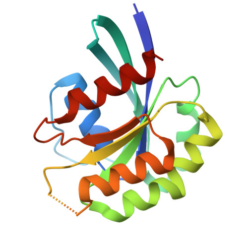
7XQV

The complex of nanobody Rh57 binding to GTP-bound RhoA active form


Experimental Data Snapshot

  • Method: X-RAY DIFFRACTION
  • Resolution: 2.76 Å
  • R-Value Free: 0.270 
  • R-Value Work: 0.220 
  • R-Value Observed: 0.223 

Starting Model: experimental
View more details

wwPDB Validation   3D Report Full Report


Ligand Structure Quality Assessment 


This is version 1.3 of the entry. See complete history


Literature

Structural insights into the binding of nanobody Rh57 to active RhoA-GTP.

Zhang, Y.Cheng, S.Zhong, P.Wang, Z.Liu, R.Ding, Y.

(2022) Biochem Biophys Res Commun 616: 122-128

  • DOI: https://doi.org/10.1016/j.bbrc.2022.05.084
  • Primary Citation of Related Structures:  
    7XQV

  • PubMed Abstract: 

    RhoA protein is a small GTPase that acts as a molecular switch. When bound to guanosine triphosphate (GTP), RhoA can activate several key signal pathways. Recently, nanobody Rh57 specific binding with GTP bound active RhoA was discovered and developed as a BRET biosensor without cytotoxicity. To further clarify the nanobody Rh57's mechanism of action, we co-expressed, purified, and crystallized the RhoA-Rh57 nanobody complex and solved the structure by X-ray diffraction with a resolution of 2.76 Å. The structure showed that the interaction is mainly through hydrogen bonds, salt bridges, aromatic-aromatic interactions, and hydrophobic interactions. The involved regions include CDR3 and non-hypervariable loop of Rh57, and the SWI switch loops of RhoA, respectively. The different SWI conformation of inactivated RhoA-GDP prevented the Rh57's binding. The possible explanation of Rh57 as a non-cytotoxic BRET intracellular tracer is that Rh57's binding did not overlap with downstream PRK1 and thus did not interfere with the downstream signaling pathway. Our research provides an in-depth understanding of how nanobodies recognize activated RhoA-GTP while not binding inactivated RhoA-GDP. This structural information may also provide critical information for further optimization of relevant nanobodies.


  • Organizational Affiliation

    State Key Laboratory of Genetic Engineering, School of Life Sciences, Fudan University, Shanghai, 200438, China.


Macromolecules
Find similar proteins by:  (by identity cutoff)  |  3D Structure
Entity ID: 1
MoleculeChains Sequence LengthOrganismDetailsImage
RhoA183Rattus norvegicusMutation(s): 0 
EC: 3.6.5.2
UniProt
Find proteins for P61589 (Rattus norvegicus)
Explore P61589 
Go to UniProtKB:  P61589
Entity Groups  
Sequence Clusters30% Identity50% Identity70% Identity90% Identity95% Identity100% Identity
UniProt GroupP61589
Sequence Annotations
Expand
  • Reference Sequence
Find similar proteins by:  (by identity cutoff)  |  3D Structure
Entity ID: 2
MoleculeChains Sequence LengthOrganismDetailsImage
Rh57145Camelus bactrianusMutation(s): 0 
Entity Groups  
Sequence Clusters30% Identity50% Identity70% Identity90% Identity95% Identity100% Identity
Sequence Annotations
Expand
  • Reference Sequence
Experimental Data & Validation

Experimental Data

  • Method: X-RAY DIFFRACTION
  • Resolution: 2.76 Å
  • R-Value Free: 0.270 
  • R-Value Work: 0.220 
  • R-Value Observed: 0.223 
  • Space Group: I 41 2 2
Unit Cell:
Length ( Å )Angle ( ˚ )
a = 142.03α = 90
b = 142.03β = 90
c = 69.26γ = 90
Software Package:
Software NamePurpose
PHENIXrefinement
PDB_EXTRACTdata extraction
DIALSdata reduction
DIALSdata scaling
PHENIXphasing

Structure Validation

View Full Validation Report


Ligand Structure Quality Assessment 


Entry History & Funding Information

Deposition Data


Funding OrganizationLocationGrant Number
National Science Foundation (NSF, China)China32070939
National Science Foundation (NSF, China)China82030106

Revision History  (Full details and data files)

  • Version 1.0: 2022-07-13
    Type: Initial release
  • Version 1.1: 2023-11-29
    Changes: Data collection, Refinement description
  • Version 1.2: 2024-05-08
    Changes: Database references
  • Version 1.3: 2024-10-30
    Changes: Structure summary