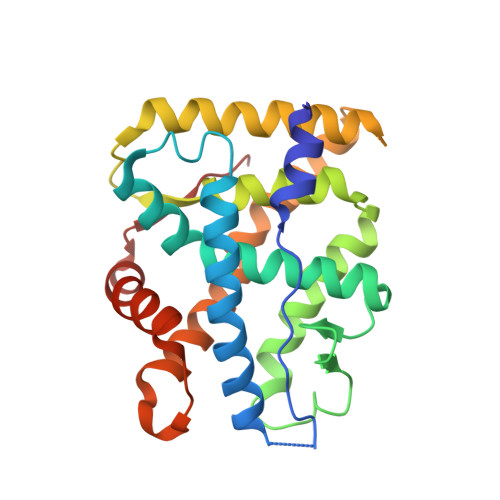
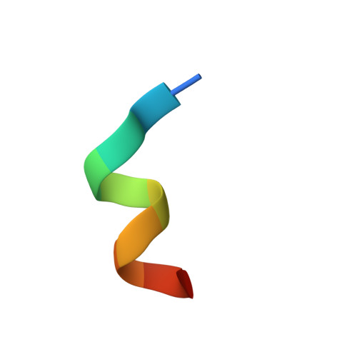
7YXO

Crystal structure of WT AncGR2-LBD bound to dexamethasone and SHP coregulator fragment


Experimental Data Snapshot

  • Method: X-RAY DIFFRACTION
  • Resolution: 2.99 Å
  • R-Value Free: 0.280 
  • R-Value Work: 0.242 
  • R-Value Observed: 0.244 

Starting Model: experimental
View more details

wwPDB Validation   3D Report Full Report


Ligand Structure Quality Assessment 


This is version 1.3 of the entry. See complete history


Literature

The multivalency of the glucocorticoid receptor ligand-binding domain explains its manifold physiological activities.

Jimenez-Panizo, A.Alegre-Marti, A.Tettey, T.T.Fettweis, G.Abella, M.Anton, R.Johnson, T.A.Kim, S.Schiltz, R.L.Nunez-Barrios, I.Font-Diaz, J.Caelles, C.Valledor, A.F.Perez, P.Rojas, A.M.Fernandez-Recio, J.Presman, D.M.Hager, G.L.Fuentes-Prior, P.Estebanez-Perpina, E.

(2022) Nucleic Acids Res 50: 13063-13082

  • DOI: https://doi.org/10.1093/nar/gkac1119
  • Primary Citation of Related Structures:  
    7YXC, 7YXD, 7YXN, 7YXO, 7YXP, 7YXR

  • PubMed Abstract: 

    The glucocorticoid receptor (GR) is a ubiquitously expressed transcription factor that controls metabolic and homeostatic processes essential for life. Although numerous crystal structures of the GR ligand-binding domain (GR-LBD) have been reported, the functional oligomeric state of the full-length receptor, which is essential for its transcriptional activity, remains disputed. Here we present five new crystal structures of agonist-bound GR-LBD, along with a thorough analysis of previous structural work. We identify four distinct homodimerization interfaces on the GR-LBD surface, which can associate into 20 topologically different homodimers. Biologically relevant homodimers were identified by studying a battery of GR point mutants including crosslinking assays in solution, quantitative fluorescence microscopy in living cells, and transcriptomic analyses. Our results highlight the relevance of non-canonical dimerization modes for GR, especially of contacts made by loop L1-3 residues such as Tyr545. Our work illustrates the unique flexibility of GR's LBD and suggests different dimeric conformations within cells. In addition, we unveil pathophysiologically relevant quaternary assemblies of the receptor with important implications for glucocorticoid action and drug design.


  • Organizational Affiliation

    Department of Biochemistry and Molecular Biomedicine, Faculty of Biology, University of Barcelona (UB), 08028 Barcelona, Spain.


Macromolecules
Find similar proteins by:  (by identity cutoff)  |  3D Structure
Entity ID: 1
MoleculeChains Sequence LengthOrganismDetailsImage
Ancestral Glucocorticoid Receptor2
A, C, E
248unidentifiedMutation(s): 0 
UniProt
Find proteins for A0A1X8XLE9 (unidentified)
Explore A0A1X8XLE9 
Go to UniProtKB:  A0A1X8XLE9
Entity Groups  
Sequence Clusters30% Identity50% Identity70% Identity90% Identity95% Identity100% Identity
UniProt GroupA0A1X8XLE9
Sequence Annotations
Expand
  • Reference Sequence

Find similar proteins by:  Sequence   |   3D Structure  

Entity ID: 2
MoleculeChains Sequence LengthOrganismDetailsImage
SHP NR Box 1 Peptide
B, D, F
11Homo sapiensMutation(s): 0 
UniProt & NIH Common Fund Data Resources
Find proteins for Q15466 (Homo sapiens)
Explore Q15466 
Go to UniProtKB:  Q15466
PHAROS:  Q15466
GTEx:  ENSG00000131910 
Entity Groups  
Sequence Clusters30% Identity50% Identity70% Identity90% Identity95% Identity100% Identity
UniProt GroupQ15466
Sequence Annotations
Expand
  • Reference Sequence
Small Molecules
Ligands 3 Unique
IDChains Name / Formula / InChI Key2D Diagram3D Interactions
CPS
Query on CPS

Download Ideal Coordinates CCD File 
H [auth A],
K [auth C]
3-[(3-CHOLAMIDOPROPYL)DIMETHYLAMMONIO]-1-PROPANESULFONATE
C32 H58 N2 O7 S
UMCMPZBLKLEWAF-BCTGSCMUSA-N
DEX (Subject of Investigation/LOI)
Query on DEX

Download Ideal Coordinates CCD File 
G [auth A],
J [auth C],
N [auth E]
DEXAMETHASONE
C22 H29 F O5
UREBDLICKHMUKA-CXSFZGCWSA-N
SO4
Query on SO4

Download Ideal Coordinates CCD File 
I [auth A],
L [auth C],
M [auth C]
SULFATE ION
O4 S
QAOWNCQODCNURD-UHFFFAOYSA-L
Experimental Data & Validation

Experimental Data

  • Method: X-RAY DIFFRACTION
  • Resolution: 2.99 Å
  • R-Value Free: 0.280 
  • R-Value Work: 0.242 
  • R-Value Observed: 0.244 
  • Space Group: I 41 2 2
Unit Cell:
Length ( Å )Angle ( ˚ )
a = 180.2α = 90
b = 180.2β = 90
c = 169.18γ = 90
Software Package:
Software NamePurpose
REFMACrefinement
PDB_EXTRACTdata extraction
MOSFLMdata reduction
SCALAdata scaling
MOLREPphasing

Structure Validation

View Full Validation Report


Ligand Structure Quality Assessment 


Entry History & Funding Information

Deposition Data


Funding OrganizationLocationGrant Number
Other governmentSpainBFU-Retos2017-86906-R
Other governmentSpainSAF2017-71878-REDT
Other governmentSpainSAF2015-71878-REDT
Other governmentSpainRTI2018-101500-B-I00

Revision History  (Full details and data files)

  • Version 1.0: 2022-12-07
    Type: Initial release
  • Version 1.1: 2022-12-14
    Changes: Database references
  • Version 1.2: 2023-01-18
    Changes: Database references
  • Version 1.3: 2024-01-31
    Changes: Data collection, Refinement description