7Z2B | pdb_00007z2b

P. berghei kinesin-8B motor domain in AMPPNP state bound to tubulin dimer


Experimental Data Snapshot

  • Method: ELECTRON MICROSCOPY
  • Resolution: 3.30 Å
  • Aggregation State: FILAMENT 
  • Reconstruction Method: SINGLE PARTICLE 

wwPDB Validation 3D Report Full Report

Validation slider image for 7Z2B

This is version 1.2 of the entry. See complete history

Literature

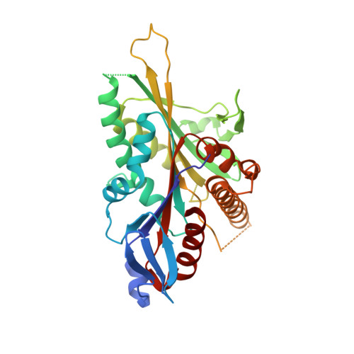
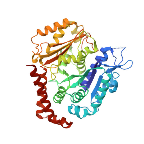
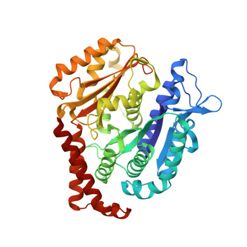
Mechanochemical tuning of a kinesin motor essential for malaria parasite transmission.

Liu, T.Shilliday, F.Cook, A.D.Zeeshan, M.Brady, D.Tewari, R.Sutherland, C.J.Roberts, A.J.Moores, C.A.

(2022) Nat Commun 13: 6988-6988

  • DOI: https://doi.org/10.1038/s41467-022-34710-x
  • Primary Citation Related Structures: 
    7Z2A, 7Z2B, 7Z2C

  • PubMed Abstract: 

    Plasmodium species cause malaria and kill hundreds of thousands annually. The microtubule-based motor kinesin-8B is required for development of the flagellated Plasmodium male gamete, and its absence completely blocks parasite transmission. To understand the molecular basis of kinesin-8B's essential role, we characterised the in vitro properties of kinesin-8B motor domains from P. berghei and P. falciparum. Both motors drive ATP-dependent microtubule gliding, but also catalyse ATP-dependent microtubule depolymerisation. We determined these motors' microtubule-bound structures using cryo-electron microscopy, which showed very similar modes of microtubule interaction in which Plasmodium-distinct sequences at the microtubule-kinesin interface influence motor function. Intriguingly however, P. berghei kinesin-8B exhibits a non-canonical structural response to ATP analogue binding such that neck linker docking is not induced. Nevertheless, the neck linker region is required for motility and depolymerisation activities of these motors. These data suggest that the mechanochemistry of Plasmodium kinesin-8Bs is functionally tuned to support flagella formation.


  • Organizational Affiliation
    • Institute of Structural and Molecular Biology, Birkbeck College, London, WC1E 7HX, UK.

Macromolecule Content 

  • Total Structure Weight: 135.68 kDa 
  • Atom Count: 9,124 
  • Modeled Residue Count: 1,162 
  • Deposited Residue Count: 1,195 
  • Unique protein chains: 3

Macromolecules

Find similar proteins by:|  3D Structure
Entity ID: 1
MoleculeChains  Sequence LengthOrganismDetailsImage
Kinesin-8, putativeA [auth K]341Plasmodium bergheiMutation(s): 0 
UniProt
Find proteins for A0A509AB64 (Plasmodium berghei (strain Anka))
Explore A0A509AB64 
Go to UniProtKB:  A0A509AB64
Entity Groups
Sequence Clusters30% Identity50% Identity70% Identity90% Identity95% Identity100% Identity
UniProt GroupA0A509AB64
Sequence Annotations
Expand
Reference Sequence
Find similar proteins by:|  3D Structure
Entity ID: 2
MoleculeChains  Sequence LengthOrganismDetailsImage
Detyrosinated tubulin alpha-1B chainB [auth A]428Sus scrofaMutation(s): 0 
EC: 3.6.5
UniProt
Find proteins for Q2XVP4 (Sus scrofa)
Explore Q2XVP4 
Go to UniProtKB:  Q2XVP4
Entity Groups
Sequence Clusters30% Identity50% Identity70% Identity90% Identity95% Identity100% Identity
UniProt GroupQ2XVP4
Sequence Annotations
Expand
Reference Sequence
Find similar proteins by:|  3D Structure
Entity ID: 3
MoleculeChains  Sequence LengthOrganismDetailsImage
Tubulin beta chainC [auth H]426Sus scrofaMutation(s): 0 
UniProt
Find proteins for P02554 (Sus scrofa)
Explore P02554 
Go to UniProtKB:  P02554
Entity Groups
Sequence Clusters30% Identity50% Identity70% Identity90% Identity95% Identity100% Identity
UniProt GroupP02554
Sequence Annotations
Expand
Reference Sequence

Small Molecules

Ligands 4 Unique
IDChains Name / Formula / InChI Key2D Diagram3D Interactions
GTP

Query on GTP


Download:Ideal Coordinates CCD File
F [auth A]GUANOSINE-5'-TRIPHOSPHATE
C10 H16 N5 O14 P3
XKMLYUALXHKNFT-UUOKFMHZSA-N
G2P
(Subject of Investigation/LOI)

Query on G2P


Download:Ideal Coordinates CCD File
H
PHOSPHOMETHYLPHOSPHONIC ACID GUANYLATE ESTER
C11 H18 N5 O13 P3
GXTIEXDFEKYVGY-KQYNXXCUSA-N
ANP

Query on ANP


Download:Ideal Coordinates CCD File
D [auth K]PHOSPHOAMINOPHOSPHONIC ACID-ADENYLATE ESTER
C10 H17 N6 O12 P3
PVKSNHVPLWYQGJ-KQYNXXCUSA-N
MG

Query on MG


Download:Ideal Coordinates CCD File
E [auth K],
G [auth A],
I [auth H]
MAGNESIUM ION
Mg
JLVVSXFLKOJNIY-UHFFFAOYSA-N

Experimental Data & Validation

Experimental Data

  • Method: ELECTRON MICROSCOPY
  • Resolution: 3.30 Å
  • Aggregation State: FILAMENT 
  • Reconstruction Method: SINGLE PARTICLE 

Structure Validation

View Full Validation Report


Entry History 

& Funding Information

Deposition Data


Funding OrganizationLocationGrant Number
Biotechnology and Biological Sciences Research Council (BBSRC)United Kingdom--
Wellcome TrustUnited Kingdom--

Revision History  (Full details and data files)

  • Version 1.0: 2022-11-16
    Type: Initial release
  • Version 1.1: 2022-11-30
    Changes: Database references
  • Version 1.2: 2024-07-17
    Changes: Data collection