7Z9P

The novel DNA binding mechanism of ridinilazole, a precision Clostridiodes difficile antibiotic


Experimental Data Snapshot

  • Method: X-RAY DIFFRACTION
  • Resolution: 2.20 Å
  • R-Value Free: 0.241 
  • R-Value Work: 0.224 
  • R-Value Observed: 0.225 

wwPDB Validation   3D Report Full Report


Ligand Structure Quality Assessment 


This is version 1.3 of the entry. See complete history


Literature

The Novel DNA Binding Mechanism of Ridinilazole, a Precision Clostridiodes difficile Antibiotic.

Mason, C.S.Avis, T.Hu, C.Nagalingam, N.Mudaliar, M.Coward, C.Begum, K.Gajewski, K.Alam, M.J.Basseres, E.Moss, S.Reich, S.Duperchy, E.Fox, K.R.Garey, K.W.Powell, D.J.

(2023) Antimicrob Agents Chemother 67: e0156322-e0156322

  • DOI: https://doi.org/10.1128/aac.01563-22
  • Primary Citation of Related Structures:  
    7Z9P

  • PubMed Abstract: 

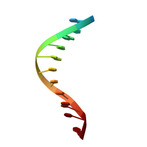
    Clostridioides difficile infection (CDI) causes substantial morbidity and mortality worldwide with limited antibiotic treatment options. Ridinilazole is a precision bisbenzimidazole antibiotic being developed to treat CDI and reduce unacceptably high rates of infection recurrence in patients. Although in late clinical development, the precise mechanism of action by which ridinilazole elicits its bactericidal activity has remained elusive. Here, we present conclusive biochemical and structural data to demonstrate that ridinilazole has a primary DNA binding mechanism, with a co-complex structure confirming binding to the DNA minor groove. Additional RNA-seq data indicated early pleiotropic changes to transcription, with broad effects on multiple C. difficile compartments and significant effects on energy generation pathways particularly. DNA binding and genomic localization was confirmed through confocal microscopy utilizing the intrinsic fluorescence of ridinilazole upon DNA binding. As such, ridinilazole has the potential to be the first antibiotic approved with a DNA minor groove binding mechanism of action.


  • Organizational Affiliation

    Summit Therapeutics, Cambridge, United Kingdom.


Macromolecules

Find similar nucleic acids by:  Sequence   |   3D Structure  

Entity ID: 1
MoleculeChains LengthOrganismImage
DNA (5'-D(P*GP*CP*GP*AP*AP*TP*TP*CP*GP*CP*G*(RID))-3')
A, B, C, D, E
A, B, C, D, E, F
12Clostridioides difficile
Sequence Annotations
Expand
  • Reference Sequence
Small Molecules
Ligands 4 Unique
IDChains Name / Formula / InChI Key2D Diagram3D Interactions
IK7 (Subject of Investigation/LOI)
Query on IK7

Download Ideal Coordinates CCD File 
G [auth A],
O [auth D],
P [auth E]
Ridinilazole
C24 H16 N6
UHQFBTAJFNVZIV-UHFFFAOYSA-N
SO4
Query on SO4

Download Ideal Coordinates CCD File 
H [auth A],
J [auth C],
K [auth C],
L [auth C],
Q [auth E]
SULFATE ION
O4 S
QAOWNCQODCNURD-UHFFFAOYSA-L
ZN
Query on ZN

Download Ideal Coordinates CCD File 
M [auth C],
N [auth C],
R [auth F]
ZINC ION
Zn
PTFCDOFLOPIGGS-UHFFFAOYSA-N
NA
Query on NA

Download Ideal Coordinates CCD File 
I [auth A]SODIUM ION
Na
FKNQFGJONOIPTF-UHFFFAOYSA-N
Experimental Data & Validation

Experimental Data

  • Method: X-RAY DIFFRACTION
  • Resolution: 2.20 Å
  • R-Value Free: 0.241 
  • R-Value Work: 0.224 
  • R-Value Observed: 0.225 
  • Space Group: P 1
Unit Cell:
Length ( Å )Angle ( ˚ )
a = 38.576α = 60.78
b = 40.32β = 87.31
c = 40.577γ = 61.62
Software Package:
Software NamePurpose
REFMACrefinement
DIALSdata reduction
Aimlessdata scaling
PHASERphasing

Structure Validation

View Full Validation Report


Ligand Structure Quality Assessment 


Entry History & Funding Information

Deposition Data


Funding OrganizationLocationGrant Number
Not funded--

Revision History  (Full details and data files)

  • Version 1.0: 2023-03-29
    Type: Initial release
  • Version 1.1: 2023-05-03
    Changes: Database references
  • Version 1.2: 2023-05-31
    Changes: Database references
  • Version 1.3: 2024-02-07
    Changes: Data collection, Refinement description