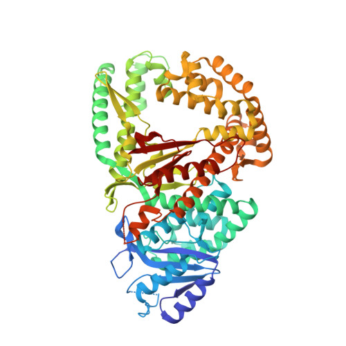
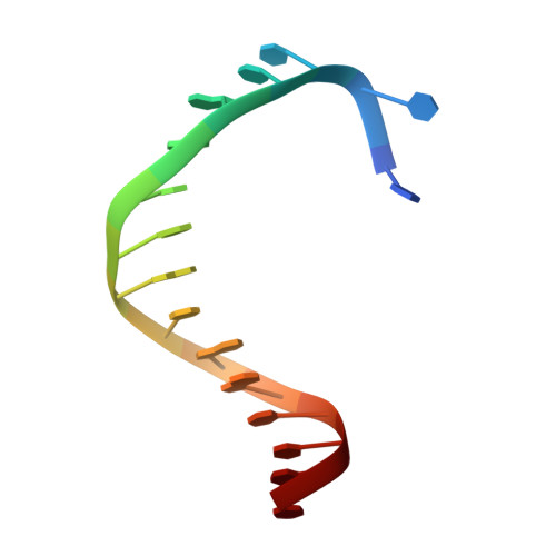
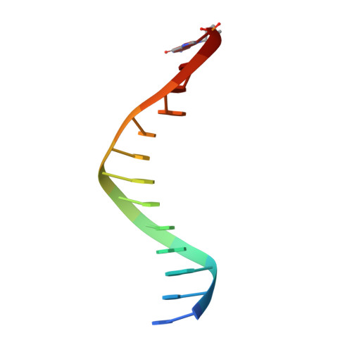
RCSB PDB - 7ZX1: Crystal structure of Pol theta polymerase domain in complex with compound 22

 7ZX1

Crystal structure of Pol theta polymerase domain in complex with compound 22


Experimental Data Snapshot

  • Method: X-RAY DIFFRACTION
  • Resolution: 2.83 Å
  • R-Value Free: 0.260 
  • R-Value Work: 0.228 

Starting Model: experimental
View more details

wwPDB Validation   3D Report Full Report


Ligand Structure Quality Assessment 

Created with Raphaël 2.3.0Worse 01 BetterLigand structure goodness of fit to experimental dataBest fitted DG3Click on this verticalbar to view detailsBest fitted K8IClick on this verticalbar to view details

This is version 1.3 of the entry. See complete history


Literature

Discovery, Characterization, and Structure-Based Optimization of Small-Molecule In Vitro and In Vivo Probes for Human DNA Polymerase Theta.

Stockley, M.L.Ferdinand, A.Benedetti, G.Blencowe, P.Boyd, S.M.Calder, M.Charles, M.D.Edwardes, L.V.Ekwuru, T.Finch, H.Galbiati, A.Geo, L.Grande, D.Grinkevich, V.Holliday, N.D.Krajewski, W.W.MacDonald, E.Majithiya, J.B.McCarron, H.McWhirter, C.L.Patel, V.Pedder, C.Rajendra, E.Ranzani, M.Rigoreau, L.J.M.Robinson, H.M.R.Schaedler, T.Sirina, J.Smith, G.C.M.Swarbrick, M.E.Turnbull, A.P.Willis, S.Heald, R.A.

(2022) J Med Chem 65: 13879-13891

  • DOI: https://doi.org/10.1021/acs.jmedchem.2c01142
  • Primary Citation of Related Structures:  
    7ZUS, 7ZX0, 7ZX1

  • PubMed Abstract: 

    Human DNA polymerase theta (Polθ), which is essential for microhomology-mediated DNA double strand break repair, has been proposed as an attractive target for the treatment of BRCA deficient and other DNA repair pathway defective cancers. As previously reported, we recently identified the first selective small molecule Polθ in vitro probe, 22 (ART558), which recapitulates the phenotype of Polθ loss, and in vivo probe, 43 (ART812), which is efficacious in a model of PARP inhibitor resistant TNBC in vivo. Here we describe the discovery, biochemical and biophysical characterization of these probes including small molecule ligand co-crystal structures with Polθ. The crystallographic data provides a basis for understanding the unique mechanism of inhibition of these compounds which is dependent on stabilization of a "closed" enzyme conformation. Additionally, the structural biology platform provided a basis for rational optimization based primarily on reduced ligand conformational flexibility.


  • Organizational Affiliation

    Artios Pharma Ltd., B940, Babraham Research Campus, CambridgeCB22 3FH, U. K.


Macromolecules

Find similar proteins by:  (by identity cutoff)  |  3D Structure
Entity ID: 1
MoleculeChains Sequence LengthOrganismDetailsImage
DNA polymerase theta726Homo sapiensMutation(s): 1 
Gene Names: POLQPOLH
EC: 2.7.7.7 (PDB Primary Data), 3.6.4.12 (UniProt)
UniProt & NIH Common Fund Data Resources
Find proteins for O75417 (Homo sapiens)
Explore O75417 
Go to UniProtKB:  O75417
PHAROS:  O75417
GTEx:  ENSG00000051341 
Entity Groups  
Sequence Clusters30% Identity50% Identity70% Identity90% Identity95% Identity100% Identity
UniProt GroupO75417
Sequence Annotations
Expand
  • Reference Sequence
Small Molecules
Ligands 3 Unique
IDChains Name / Formula / InChI Key2D Diagram3D Interactions
DG3 (Subject of Investigation/LOI)
Query on DG3

Download Ideal Coordinates CCD File 
BA [auth DDD]
EA [auth EEE]
GA [auth FFF]
HA [auth KKK]
T [auth AAA]
2'-3'-DIDEOXYGUANOSINE-5'-TRIPHOSPHATE
C10 H16 N5 O12 P3
HDRRAMINWIWTNU-NTSWFWBYSA-N
K8I (Subject of Investigation/LOI)
Query on K8I

Download Ideal Coordinates CCD File 
CA [auth DDD],
U [auth AAA],
X [auth BBB],
Z [auth CCC]
(2~{S},3~{R})-1-[3-cyano-6-methyl-4-(trifluoromethyl)pyridin-2-yl]-~{N}-methyl-~{N}-(3-methylphenyl)-3-oxidanyl-pyrrolidine-2-carboxamide
C21 H21 F3 N4 O2
YHMDHAMZFMNMTF-MSOLQXFVSA-N
MG
Query on MG

Download Ideal Coordinates CCD File 
AA [auth DDD]
DA [auth EEE]
FA [auth FFF]
S [auth AAA]
V [auth BBB]
MAGNESIUM ION
Mg
JLVVSXFLKOJNIY-UHFFFAOYSA-N
Binding Affinity Annotations 
IDSourceBinding Affinity
K8I BindingDB:  7ZX1 Kd: min: 4.8, max: 6.4 (nM) from 2 assay(s)
IC50: min: 7.9, max: 1.20e+4 (nM) from 2 assay(s)
Experimental Data & Validation

Experimental Data

  • Method: X-RAY DIFFRACTION
  • Resolution: 2.83 Å
  • R-Value Free: 0.260 
  • R-Value Work: 0.228 
  • Space Group: P 1 21 1
Unit Cell:
Length ( Å )Angle ( ˚ )
a = 59.589α = 90
b = 172.292β = 91.217
c = 289.209γ = 90
Software Package:
Software NamePurpose
REFMACrefinement
xia2data reduction
xia2data scaling
PHASERphasing
Cootmodel building

Structure Validation

View Full Validation Report


Ligand Structure Quality Assessment 

Created with Raphaël 2.3.0Worse 01 BetterLigand structure goodness of fit to experimental dataBest fitted DG3Click on this verticalbar to view detailsBest fitted K8IClick on this verticalbar to view details

Entry History & Funding Information

Deposition Data


Funding OrganizationLocationGrant Number
Other private--

Revision History  (Full details and data files)

  • Version 1.0: 2022-10-12
    Type: Initial release
  • Version 1.1: 2022-10-19
    Changes: Database references
  • Version 1.2: 2022-11-09
    Changes: Database references
  • Version 1.3: 2024-01-31
    Changes: Data collection, Refinement description