7CIO | pdb_00007cio

Molecular interactions of cytoplasmic region of CTLA-4 with SH2 domains of PI3-kinase


Experimental Data Snapshot

  • Method: X-RAY DIFFRACTION
  • Resolution: 1.10 Å
  • R-Value Free: 
    0.179 (Depositor), 0.179 (DCC) 
  • R-Value Work: 
    0.154 (Depositor), 0.154 (DCC) 
  • R-Value Observed: 
    0.155 (Depositor) 

Starting Model: experimental
View more details

wwPDB Validation 3D Report Full Report

Validation slider image for 7CIO

This is version 1.3 of the entry. See complete history

Literature

Molecular interactions of the CTLA-4 cytoplasmic region with the phosphoinositide 3-kinase SH2 domains.

Iiyama, M.Numoto, N.Ogawa, S.Kuroda, M.Morii, H.Abe, R.Ito, N.Oda, M.

(2021) Mol Immunol 131: 51-59

  • DOI: https://doi.org/10.1016/j.molimm.2020.12.002
  • Primary Citation Related Structures: 
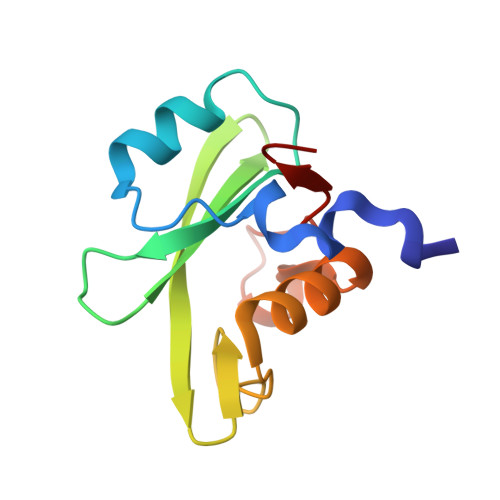
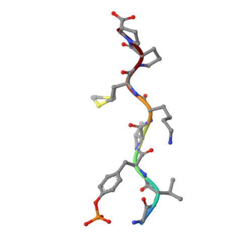
    7CIO

  • PubMed Abstract: 

    During T-cell regulation, T-cell receptors and CD28 lead to signaling activation, while T-lymphocyte antigen 4 (CTLA-4) is known to lead to downregulation, similar to programmed cell death-1 (PD-1). In the cytoplasmic tails of CD28 and CTLA-4, phosphoinositide 3-kinase (PI3K) binds to the consensus sequence including phosphotyrosine via SH2 domains, N- and C-terminal SH2 domains (nSH2 and cSH2), of its regulatory subunit, p85. In this study, we determined the crystal structure of a CTLA-4-derived phosphopeptide in complex with a Cys-substituted mutant of cSH2, C656S/C659V/C670L, at a 1.1 Å resolution. Phosphotyrosine of the bound peptide is tightly accommodated by the residues Arg631, Arg649, Ser651, and Ser652, similar to the cSH2 wild-type recognition mode of CD28, as reported previously. Upon the Cys mutation, the cSH2 thermal stability increased while the CTLA-4 binding affinity slightly changed. The binding experiments also showed that the binding affinity of CTLA-4 by cSH2 was approximately two orders of magnitude lower than that of CD28. Similar to CD28 binding, the CTLA-4 binding affinity of nSH2 was lower than that of cSH2. The complex structure of nSH2 and CTLA-4 was modeled, and compared with the crystal structure of cSH2 mutant and CTLA-4. The difference in the binding affinity between CD28 and CTLA-4, along with the difference between nSH2 and cSH2, could be explained by the 3D structures, which would be closely correlated with the respective T-cell signaling.


  • Organizational Affiliation
    • Graduate School of Life and Environmental Sciences, Kyoto Prefectural University, 1-5 Hangi-cho, Shimogamo, Sakyo-ku, Kyoto 606-8522, Japan.

Macromolecule Content 

  • Total Structure Weight: 13.15 kDa 
  • Atom Count: 1,126 
  • Modeled Residue Count: 117 
  • Deposited Residue Count: 117 
  • Unique protein chains: 2

Macromolecules

Find similar proteins by:|  3D Structure
Entity ID: 1
MoleculeChains  Sequence LengthOrganismDetailsImage
Phosphatidylinositol 3-kinase regulatory subunit alpha109Homo sapiensMutation(s): 3 
Gene Names: PIK3R1GRB1
UniProt & NIH Common Fund Data Resources
Find proteins for P27986 (Homo sapiens)
Explore P27986 
Go to UniProtKB:  P27986
PHAROS:  P27986
GTEx:  ENSG00000145675 
Entity Groups
Sequence Clusters30% Identity50% Identity70% Identity90% Identity95% Identity100% Identity
UniProt GroupP27986
Sequence Annotations
Expand
Reference Sequence
Find similar proteins by:  Sequence   |   3D Structure  
Entity ID: 2
MoleculeChains  Sequence LengthOrganismDetailsImage
Cytotoxic T-lymphocyte protein 48Homo sapiensMutation(s): 0 
UniProt & NIH Common Fund Data Resources
Find proteins for P16410 (Homo sapiens)
Explore P16410 
Go to UniProtKB:  P16410
PHAROS:  P16410
GTEx:  ENSG00000163599 
Entity Groups
UniProt GroupP16410
Sequence Annotations
Expand
Reference Sequence

Small Molecules

Modified Residues  1 Unique
IDChains TypeFormula2D DiagramParent
PTR
Query on PTR
B
L-PEPTIDE LINKINGC9 H12 N O6 PTYR

Experimental Data & Validation

Experimental Data

  • Method: X-RAY DIFFRACTION
  • Resolution: 1.10 Å
  • R-Value Free:  0.179 (Depositor), 0.179 (DCC) 
  • R-Value Work:  0.154 (Depositor), 0.154 (DCC) 
  • R-Value Observed: 0.155 (Depositor) 
Space Group: P 21 21 21
Unit Cell:
Length ( Å )Angle ( ˚ )
a = 41.221α = 90
b = 41.563β = 90
c = 59.628γ = 90
Software Package:
Software NamePurpose
PHENIXrefinement
XDSdata reduction
XDSdata scaling
PHASERphasing

Structure Validation

View Full Validation Report


Entry History 

Deposition Data

Revision History  (Full details and data files)

  • Version 1.0: 2020-12-09
    Type: Initial release
  • Version 1.1: 2021-03-17
    Changes: Database references
  • Version 1.2: 2023-11-29
    Changes: Data collection, Database references, Refinement description
  • Version 1.3: 2024-10-30
    Changes: Structure summary