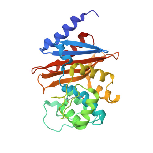
7RPF

X-ray crystal structure of OXA-24/40 in complex with doripenem


Experimental Data Snapshot

  • Method: X-RAY DIFFRACTION
  • Resolution: 1.90 Å
  • R-Value Free: 0.205 
  • R-Value Work: 0.172 
  • R-Value Observed: 0.173 

Starting Model: experimental
View more details

wwPDB Validation   3D Report Full Report


Ligand Structure Quality Assessment 


This is version 1.3 of the entry. See complete history


Literature

Conformational flexibility in carbapenem hydrolysis drives substrate specificity of the class D carbapenemase OXA-24/40.

Mitchell, J.M.June, C.M.Baggett, V.L.Lowe, B.C.Ruble, J.F.Bonomo, R.A.Leonard, D.A.Powers, R.A.

(2022) J Biol Chem 298: 102127-102127

  • DOI: https://doi.org/10.1016/j.jbc.2022.102127
  • Primary Citation of Related Structures:  
    7RP8, 7RP9, 7RPA, 7RPB, 7RPC, 7RPD, 7RPE, 7RPF, 7RPG

  • PubMed Abstract: 

    The evolution of multidrug resistance in Acinetobacter spp. increases the risk of our best antibiotics losing their efficacy. From a clinical perspective, the carbapenem-hydrolyzing class D β-lactamase subfamily present in Acinetobacter spp. is particularly concerning because of its ability to confer resistance to carbapenems. The kinetic profiles of class D β-lactamases exhibit variability in carbapenem hydrolysis, suggesting functional differences. To better understand the structure-function relationship between the carbapenem-hydrolyzing class D β-lactamase OXA-24/40 found in Acinetobacter baumannii and carbapenem substrates, we analyzed steady-state kinetics with the carbapenem antibiotics meropenem and ertapenem and determined the structures of complexes of OXA-24/40 bound to imipenem, meropenem, doripenem, and ertapenem, as well as the expanded-spectrum cephalosporin cefotaxime, using X-ray crystallography. We show that OXA-24/40 exhibits a preference for ertapenem compared with meropenem, imipenem, and doripenem, with an increase in catalytic efficiency of up to fourfold. We suggest that superposition of the nine OXA-24/40 complexes will better inform future inhibitor design efforts by providing insight into the complicated and varying ways in which carbapenems are selected and bound by class D β-lactamases.


  • Organizational Affiliation

    Department of Chemistry, Grand Valley State University, Allendale, Michigan, USA.


Macromolecules
Find similar proteins by:  (by identity cutoff)  |  3D Structure
Entity ID: 1
MoleculeChains Sequence LengthOrganismDetailsImage
Beta-lactamase245Acinetobacter baumanniiMutation(s): 0 
Gene Names: blaOXA-33bla-OXA-40blaOXA-24blaOXA-40oxa-24oxa40SI89_16690
EC: 3.5.2.6
UniProt
Find proteins for Q8RLA6 (Acinetobacter baumannii)
Explore Q8RLA6 
Go to UniProtKB:  Q8RLA6
Entity Groups  
Sequence Clusters30% Identity50% Identity70% Identity90% Identity95% Identity100% Identity
UniProt GroupQ8RLA6
Sequence Annotations
Expand
  • Reference Sequence
Small Molecules
Ligands 4 Unique
IDChains Name / Formula / InChI Key2D Diagram3D Interactions
DRW (Subject of Investigation/LOI)
Query on DRW

Download Ideal Coordinates CCD File 
D [auth A](2S,3R,4S)-2-[(2S,3R)-3-hydroxy-1-oxobutan-2-yl]-3-methyl-4-({(3S,5S)-5-[(sulfamoylamino)methyl]pyrrolidin-3-yl}sulfanyl)-3,4-dihydro-2H-pyrrole-5-carboxylic acid
C15 H26 N4 O6 S2
RBKUPZPTQNSPAV-CFINEGTKSA-N
4J6 (Subject of Investigation/LOI)
Query on 4J6

Download Ideal Coordinates CCD File 
C [auth A](4R,5S)-5-[(2S,3R)-3-hydroxy-1-oxobutan-2-yl]-4-methyl-3-({(3S,5S)-5-[(sulfamoylamino)methyl]pyrrolidin-3-yl}sulfanyl)-4,5-dihydro-1H-pyrrole-2-carboxylic acid
C15 H26 N4 O6 S2
QJKQOMNNBOTMCR-PQTSNVLCSA-N
SO4
Query on SO4

Download Ideal Coordinates CCD File 
E [auth A],
F [auth A],
G [auth A]
SULFATE ION
O4 S
QAOWNCQODCNURD-UHFFFAOYSA-L
BCT
Query on BCT

Download Ideal Coordinates CCD File 
B [auth A]BICARBONATE ION
C H O3
BVKZGUZCCUSVTD-UHFFFAOYSA-M
Modified Residues  1 Unique
IDChains TypeFormula2D DiagramParent
KCX
Query on KCX
A
L-PEPTIDE LINKINGC7 H14 N2 O4LYS
Experimental Data & Validation

Experimental Data

  • Method: X-RAY DIFFRACTION
  • Resolution: 1.90 Å
  • R-Value Free: 0.205 
  • R-Value Work: 0.172 
  • R-Value Observed: 0.173 
  • Space Group: P 41 21 2
Unit Cell:
Length ( Å )Angle ( ˚ )
a = 102.699α = 90
b = 102.699β = 90
c = 84.468γ = 90
Software Package:
Software NamePurpose
REFMACrefinement
SCALEPACKdata scaling
PHASERphasing
PDB_EXTRACTdata extraction
HKL-2000data reduction

Structure Validation

View Full Validation Report


Ligand Structure Quality Assessment 


Entry History & Funding Information

Deposition Data


Funding OrganizationLocationGrant Number
National Institutes of Health/National Institute Of Allergy and Infectious Diseases (NIH/NIAID)United StatesR15AI094489-02
National Institutes of Health/National Institute Of Allergy and Infectious Diseases (NIH/NIAID)United StatesR01AI072219

Revision History  (Full details and data files)

  • Version 1.0: 2022-07-06
    Type: Initial release
  • Version 1.1: 2022-07-27
    Changes: Database references
  • Version 1.2: 2023-10-18
    Changes: Data collection, Refinement description
  • Version 1.3: 2023-11-15
    Changes: Data collection