8AHQ

VirD/holo-ACP5b of Streptomyces virginiae complex


Experimental Data Snapshot

  • Method: X-RAY DIFFRACTION
  • Resolution: 2.10 Å
  • R-Value Free: 0.238 
  • R-Value Work: 0.183 
  • R-Value Observed: 0.186 

wwPDB Validation   3D Report Full Report


Ligand Structure Quality Assessment 


This is version 1.1 of the entry. See complete history


Literature

Decrypting the programming of beta-methylation in virginiamycin M biosynthesis.

Collin, S.Cox, R.J.Paris, C.Jacob, C.Chagot, B.Weissman, K.J.Gruez, A.

(2023) Nat Commun 14: 1327-1327

  • DOI: https://doi.org/10.1038/s41467-023-36974-3
  • Primary Citation of Related Structures:  
    8A7Z, 8AHQ, 8AHZ, 8AIG, 8ALL

  • PubMed Abstract: 

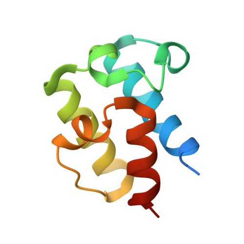
    During biosynthesis by multi-modular trans-AT polyketide synthases, polyketide structural space can be expanded by conversion of initially-formed electrophilic β-ketones into β-alkyl groups. These multi-step transformations are catalysed by 3-hydroxy-3-methylgluratryl synthase cassettes of enzymes. While mechanistic aspects of these reactions have been delineated, little information is available concerning how the cassettes select the specific polyketide intermediate(s) to target. Here we use integrative structural biology to identify the basis for substrate choice in module 5 of the virginiamycin M trans-AT polyketide synthase. Additionally, we show in vitro that module 7, at minimum, is a potential additional site for β-methylation. Indeed, analysis by HPLC-MS coupled with isotopic labelling and pathway inactivation identifies a metabolite bearing a second β-methyl at the expected position. Collectively, our results demonstrate that several control mechanisms acting in concert underpin β-branching programming. Furthermore, variations in this control - whether natural or by design - open up avenues for diversifying polyketide structures towards high-value derivatives.


  • Organizational Affiliation

    Université de Lorraine, CNRS, IMoPA, F-54000, Nancy, France.


Macromolecules
Find similar proteins by:  (by identity cutoff)  |  3D Structure
Entity ID: 1
MoleculeChains Sequence LengthOrganismDetailsImage
Enoyl-CoA hydrataseA,
C [auth B]
250Streptomyces virginiaeMutation(s): 0 
Gene Names: virD
UniProt
Find proteins for A4PHM7 (Streptomyces virginiae)
Explore A4PHM7 
Go to UniProtKB:  A4PHM7
Entity Groups  
Sequence Clusters30% Identity50% Identity70% Identity90% Identity95% Identity100% Identity
UniProt GroupA4PHM7
Sequence Annotations
Expand
  • Reference Sequence
Find similar proteins by:  (by identity cutoff)  |  3D Structure
Entity ID: 2
MoleculeChains Sequence LengthOrganismDetailsImage
Hybrid polyketide synthase-non ribosomal peptide synthetaseB [auth C],
D
88Streptomyces virginiaeMutation(s): 0 
Gene Names: virA
UniProt
Find proteins for A4PHN0 (Streptomyces virginiae)
Explore A4PHN0 
Go to UniProtKB:  A4PHN0
Entity Groups  
Sequence Clusters30% Identity50% Identity70% Identity90% Identity95% Identity100% Identity
UniProt GroupA4PHN0
Sequence Annotations
Expand
  • Reference Sequence
Small Molecules
Ligands 4 Unique
IDChains Name / Formula / InChI Key2D Diagram3D Interactions
PNS (Subject of Investigation/LOI)
Query on PNS

Download Ideal Coordinates CCD File 
L [auth C],
O [auth D]
4'-PHOSPHOPANTETHEINE
C11 H23 N2 O7 P S
JDMUPRLRUUMCTL-VIFPVBQESA-N
EDO
Query on EDO

Download Ideal Coordinates CCD File 
E [auth A]
F [auth A]
G [auth A]
H [auth A]
I [auth A]
E [auth A],
F [auth A],
G [auth A],
H [auth A],
I [auth A],
M [auth B]
1,2-ETHANEDIOL
C2 H6 O2
LYCAIKOWRPUZTN-UHFFFAOYSA-N
CL
Query on CL

Download Ideal Coordinates CCD File 
J [auth A],
N [auth B]
CHLORIDE ION
Cl
VEXZGXHMUGYJMC-UHFFFAOYSA-M
NA
Query on NA

Download Ideal Coordinates CCD File 
K [auth A]SODIUM ION
Na
FKNQFGJONOIPTF-UHFFFAOYSA-N
Modified Residues  1 Unique
IDChains TypeFormula2D DiagramParent
MSE
Query on MSE
A,
C [auth B]
L-PEPTIDE LINKINGC5 H11 N O2 SeMET
Experimental Data & Validation

Experimental Data

  • Method: X-RAY DIFFRACTION
  • Resolution: 2.10 Å
  • R-Value Free: 0.238 
  • R-Value Work: 0.183 
  • R-Value Observed: 0.186 
  • Space Group: H 3
Unit Cell:
Length ( Å )Angle ( ˚ )
a = 144.878α = 90
b = 144.878β = 90
c = 88.781γ = 120
Software Package:
Software NamePurpose
XDSdata reduction
Aimlessdata scaling
MOLREPphasing
REFMACrefinement
PDB_EXTRACTdata extraction

Structure Validation

View Full Validation Report


Ligand Structure Quality Assessment 


Entry History & Funding Information

Deposition Data


Funding OrganizationLocationGrant Number
Agence Nationale de la Recherche (ANR)FranceANR-11-JSV8-003-01
Agence Nationale de la Recherche (ANR)FranceANR-16-CE92-0006-01
Agence Nationale de la Recherche (ANR)FranceANR-20-CE93-0002-01

Revision History  (Full details and data files)

  • Version 1.0: 2023-03-15
    Type: Initial release
  • Version 1.1: 2023-03-22
    Changes: Database references