8BB4

Structure of human WDR5 and pVHL:ElonginC:ElonginB bound to PROTAC with C3 linker


Experimental Data Snapshot

  • Method: X-RAY DIFFRACTION
  • Resolution: 2.80 Å
  • R-Value Free: 0.279 
  • R-Value Work: 0.203 
  • R-Value Observed: 0.206 

Starting Model: experimental
View more details

wwPDB Validation   3D Report Full Report


Ligand Structure Quality Assessment 


This is version 1.2 of the entry. See complete history


Literature

Tracking the PROTAC degradation pathway in living cells highlights the importance of ternary complex measurement for PROTAC optimization.

Schwalm, M.P.Kramer, A.Dolle, A.Weckesser, J.Yu, X.Jin, J.Saxena, K.Knapp, S.

(2023) Cell Chem Biol 30: 753-765.e8

  • DOI: https://doi.org/10.1016/j.chembiol.2023.06.002
  • Primary Citation of Related Structures:  
    7Q2J, 8BB4, 8BB5, 8C13

  • PubMed Abstract: 

    The multi-step degradation process of PROteolysis TArgeting Chimeras (PROTACs) poses a challenge for their rational development, as the rate-limiting steps that determine PROTACs efficiency remain largely unknown. Moreover, the slow throughput of currently used endpoint assays does not allow the comprehensive analysis of larger series of PROTACs. Here, we developed cell-based assays using the NanoLuciferase and HaloTag that allow measuring PROTAC-induced degradation and ternary complex formation kinetics and stability in cells. Using PROTACs developed for the degradation of WD40 repeat domain protein 5 (WDR5), the characterization of the mode of action of these PROTACs in the early degradation cascade revealed a key role of ternary complex formation and stability. Comparing a series of ternary complex crystal structures highlighted the importance of an efficient E3-target interface for ternary complex stability. The developed assays outline a strategy for the rational optimization of PROTACs using a series of live cell assays monitoring key steps of the early PROTAC-induced degradation pathway.


  • Organizational Affiliation

    Institut für Pharmazeutische Chemie, Goethe-University Frankfurt, Biozentrum, Max-von-Laue-Str. 9, 60438 Frankfurt am Main, Germany; Structural Genomics Consortium, Goethe-University Frankfurt, Buchmann Institute for Life Sciences, Max-von-Laue-Str. 15, 60438 Frankfurt am Main, Germany.


Macromolecules
Find similar proteins by:  (by identity cutoff)  |  3D Structure
Entity ID: 1
MoleculeChains Sequence LengthOrganismDetailsImage
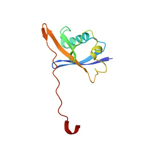
Elongin-BA [auth I]104Homo sapiensMutation(s): 0 
Gene Names: ELOBTCEB2
UniProt & NIH Common Fund Data Resources
Find proteins for Q15370 (Homo sapiens)
Explore Q15370 
Go to UniProtKB:  Q15370
PHAROS:  Q15370
GTEx:  ENSG00000103363 
Entity Groups  
Sequence Clusters30% Identity50% Identity70% Identity90% Identity95% Identity100% Identity
UniProt GroupQ15370
Sequence Annotations
Expand
  • Reference Sequence
Find similar proteins by:  (by identity cutoff)  |  3D Structure
Entity ID: 2
MoleculeChains Sequence LengthOrganismDetailsImage
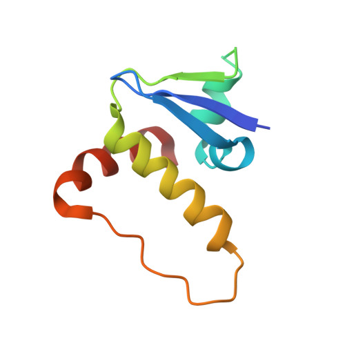
Elongin-CB [auth O]98Homo sapiensMutation(s): 0 
Gene Names: ELOCTCEB1
UniProt & NIH Common Fund Data Resources
Find proteins for Q15369 (Homo sapiens)
Explore Q15369 
Go to UniProtKB:  Q15369
PHAROS:  Q15369
GTEx:  ENSG00000154582 
Entity Groups  
Sequence Clusters30% Identity50% Identity70% Identity90% Identity95% Identity100% Identity
UniProt GroupQ15369
Sequence Annotations
Expand
  • Reference Sequence
Find similar proteins by:  (by identity cutoff)  |  3D Structure
Entity ID: 3
MoleculeChains Sequence LengthOrganismDetailsImage
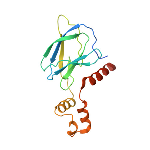
von Hippel-Lindau disease tumor suppressorC [auth P]162Homo sapiensMutation(s): 0 
Gene Names: VHL
UniProt & NIH Common Fund Data Resources
Find proteins for P40337 (Homo sapiens)
Explore P40337 
Go to UniProtKB:  P40337
PHAROS:  P40337
GTEx:  ENSG00000134086 
Entity Groups  
Sequence Clusters30% Identity50% Identity70% Identity90% Identity95% Identity100% Identity
UniProt GroupP40337
Sequence Annotations
Expand
  • Reference Sequence
Find similar proteins by:  (by identity cutoff)  |  3D Structure
Entity ID: 4
MoleculeChains Sequence LengthOrganismDetailsImage
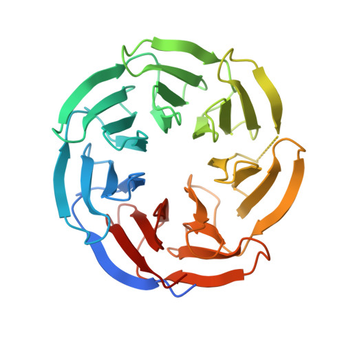
WD repeat-containing protein 5D [auth Q]321Homo sapiensMutation(s): 0 
Gene Names: WDR5BIG3
UniProt & NIH Common Fund Data Resources
Find proteins for P61964 (Homo sapiens)
Explore P61964 
Go to UniProtKB:  P61964
PHAROS:  P61964
GTEx:  ENSG00000196363 
Entity Groups  
Sequence Clusters30% Identity50% Identity70% Identity90% Identity95% Identity100% Identity
UniProt GroupP61964
Sequence Annotations
Expand
  • Reference Sequence
Small Molecules
Ligands 1 Unique
IDChains Name / Formula / InChI Key2D Diagram3D Interactions
Q3R (Subject of Investigation/LOI)
Query on Q3R

Download Ideal Coordinates CCD File 
E [auth P]~{N}-[5-[4-[[4-[[(2~{S})-3,3-dimethyl-1-[(2~{S},4~{R})-2-[[4-(4-methyl-1,3-thiazol-5-yl)phenyl]methylcarbamoyl]-4-oxidanyl-pyrrolidin-1-yl]-1-oxidanylidene-butan-2-yl]amino]-4-oxidanylidene-butyl]carbamoyl]phenyl]-2-(4-methylpiperazin-1-yl)phenyl]-6-oxidanyl-4-(trifluoromethyl)pyridine-3-carboxamide
C51 H58 F3 N9 O7 S
KFCLTULBYCCZBG-LTJJNQLXSA-N
Experimental Data & Validation

Experimental Data

  • Method: X-RAY DIFFRACTION
  • Resolution: 2.80 Å
  • R-Value Free: 0.279 
  • R-Value Work: 0.203 
  • R-Value Observed: 0.206 
  • Space Group: P 1 21 1
Unit Cell:
Length ( Å )Angle ( ˚ )
a = 46.786α = 90
b = 184.506β = 102.78
c = 47.951γ = 90
Software Package:
Software NamePurpose
Aimlessdata scaling
MOLREPphasing
REFMACrefinement
PDB_EXTRACTdata extraction
xia2data reduction

Structure Validation

View Full Validation Report


Ligand Structure Quality Assessment 


Entry History & Funding Information

Deposition Data


Funding OrganizationLocationGrant Number
The Structural Genomics Consortium (SGC)Canada--

Revision History  (Full details and data files)

  • Version 1.0: 2022-11-09
    Type: Initial release
  • Version 1.1: 2023-11-22
    Changes: Data collection, Database references
  • Version 1.2: 2024-01-31
    Changes: Refinement description