8CI8

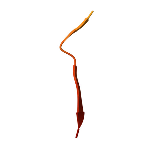
Cryo-EM structure of the Nup98(298-327) fibril


Experimental Data Snapshot

  • Method: ELECTRON MICROSCOPY
  • Resolution: 2.67 Å
  • Aggregation State: FILAMENT 
  • Reconstruction Method: HELICAL 

wwPDB Validation   3D Report Full Report


This is version 1.1 of the entry. See complete history


Literature

Impact of distinct FG nucleoporin repeats on Nup98 self-association.

Ibanez de Opakua, A.Pantoja, C.F.Cima-Omori, M.S.Dienemann, C.Zweckstetter, M.

(2024) Nat Commun 15: 3797-3797

  • DOI: https://doi.org/10.1038/s41467-024-48194-4
  • Primary Citation of Related Structures:  
    8CI8

  • PubMed Abstract: 

    Nucleoporins rich in phenylalanine/glycine (FG) residues form the permeability barrier within the nuclear pore complex and are implicated in several pathological cellular processes, including oncogenic fusion condensates. The self-association of FG-repeat proteins and interactions between FG-repeats play a critical role in these activities by forming hydrogel-like structures. Here we show that mutation of specific FG repeats of Nup98 can strongly decrease the protein's self-association capabilities. We further present a cryo-electron microscopy structure of a Nup98 peptide fibril with higher stability per residue compared with previous Nup98 fibril structures. The high-resolution structure reveals zipper-like hydrophobic patches which contain a GLFG motif and are less compatible for binding to nuclear transport receptors. The identified distinct molecular properties of different regions of the nucleoporin may contribute to spatial variations in the self-association of FG-repeats, potentially influencing transport processes through the nuclear pore.


  • Organizational Affiliation

    German Center for Neurodegenerative Diseases (DZNE), Von-Siebold-Str. 3a, Göttingen, Germany.


Macromolecules
Find similar proteins by:  (by identity cutoff)  |  3D Structure
Entity ID: 1
MoleculeChains Sequence LengthOrganismDetailsImage
Nuclear pore complex protein Nup98
A, B, C, D, E
A, B, C, D, E, F, G, H, I, J, K, L, M, N, O, P, Q, R, S, T, U, V, W, X, Y
30Homo sapiensMutation(s): 0 
EC: 3.4.21
UniProt & NIH Common Fund Data Resources
Find proteins for P52948 (Homo sapiens)
Explore P52948 
Go to UniProtKB:  P52948
PHAROS:  P52948
GTEx:  ENSG00000110713 
Entity Groups  
Sequence Clusters30% Identity50% Identity70% Identity90% Identity95% Identity100% Identity
UniProt GroupP52948
Sequence Annotations
Expand
  • Reference Sequence
Experimental Data & Validation

Experimental Data

  • Method: ELECTRON MICROSCOPY
  • Resolution: 2.67 Å
  • Aggregation State: FILAMENT 
  • Reconstruction Method: HELICAL 
EM Software:
TaskSoftware PackageVersion
MODEL REFINEMENTPHENIX
RECONSTRUCTIONRELION3.1

Structure Validation

View Full Validation Report


Entry History & Funding Information

Deposition Data


Funding OrganizationLocationGrant Number
European Research Council (ERC)European Union787679

Revision History  (Full details and data files)

  • Version 1.0: 2024-02-21
    Type: Initial release
  • Version 1.1: 2024-05-22
    Changes: Database references