8DE6

Oligomeric C9 in complex with aE11 Fab


Experimental Data Snapshot

  • Method: ELECTRON MICROSCOPY
  • Resolution: 3.20 Å
  • Aggregation State: PARTICLE 
  • Reconstruction Method: SINGLE PARTICLE 

wwPDB Validation   3D Report Full Report


This is version 1.0 of the entry. See complete history


Literature

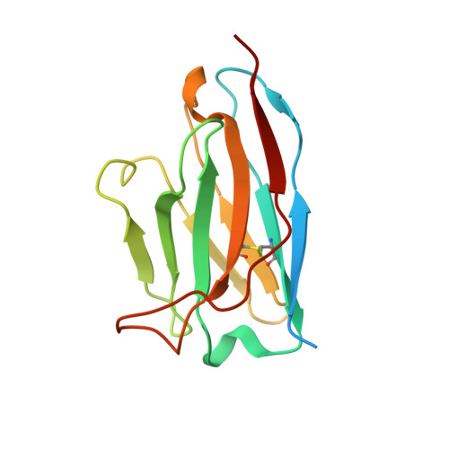
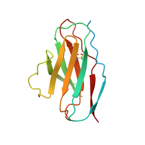
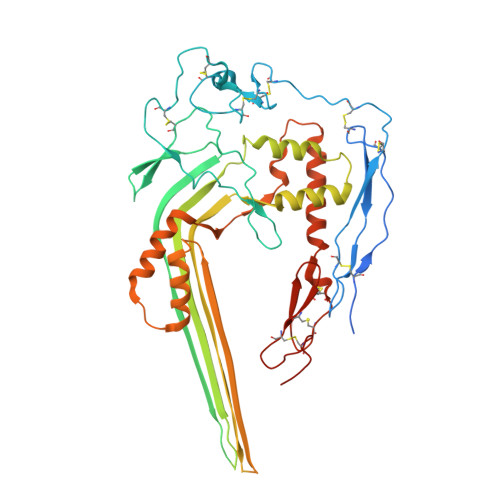
The neoepitope of the complement C5b-9 Membrane Attack Complex is formed by proximity of adjacent ancillary regions of C9.

Bayly-Jones, C.Ho, B.H.T.Lau, C.Leung, E.W.W.D'Andrea, L.Lupton, C.J.Ekkel, S.M.Venugopal, H.Whisstock, J.C.Mollnes, T.E.Spicer, B.A.Dunstone, M.A.

(2023) Commun Biol 6: 42-42

  • DOI: https://doi.org/10.1038/s42003-023-04431-y
  • Primary Citation of Related Structures:  
    8DE6

  • PubMed Abstract: 

    The Membrane Attack Complex (MAC) is responsible for forming large β-barrel channels in the membranes of pathogens, such as gram-negative bacteria. Off-target MAC assembly on endogenous tissue is associated with inflammatory diseases and cancer. Accordingly, a human C5b-9 specific antibody, aE11, has been developed that detects a neoepitope exposed in C9 when it is incorporated into the C5b-9 complex, but not present in the plasma native C9. For nearly four decades aE11 has been routinely used to study complement, MAC-related inflammation, and pathophysiology. However, the identity of C9 neoepitope remains unknown. Here, we determined the cryo-EM structure of aE11 in complex with polyC9 at 3.2 Å resolution. The aE11 binding site is formed by two separate surfaces of the oligomeric C9 periphery and is therefore a discontinuous quaternary epitope. These surfaces are contributed by portions of the adjacent TSP1, LDLRA, and MACPF domains of two neighbouring C9 protomers. By substituting key antibody interacting residues to the murine orthologue, we validated the unusual binding modality of aE11. Furthermore, aE11 can recognise a partial epitope in purified monomeric C9 in vitro, albeit weakly. Taken together, our results reveal the structural basis for MAC recognition by aE11.


  • Organizational Affiliation

    Biomedicine Discovery Institute, Department of Biochemistry and Molecular Biology, Monash University, Melbourne, VIC, Australia.


Macromolecules
Find similar proteins by:  (by identity cutoff)  |  3D Structure
Entity ID: 1
MoleculeChains Sequence LengthOrganismDetailsImage
aE11 Fab VHA [auth E],
D [auth B]
140Mus musculusMutation(s): 0 
Entity Groups  
Sequence Clusters30% Identity50% Identity70% Identity90% Identity95% Identity100% Identity
Sequence Annotations
Expand
  • Reference Sequence
Find similar proteins by:  (by identity cutoff)  |  3D Structure
Entity ID: 2
MoleculeChains Sequence LengthOrganismDetailsImage
aE11 Fab VLB [auth F],
E [auth I]
126Mus musculusMutation(s): 0 
Entity Groups  
Sequence Clusters30% Identity50% Identity70% Identity90% Identity95% Identity100% Identity
Sequence Annotations
Expand
  • Reference Sequence
Find similar proteins by:  (by identity cutoff)  |  3D Structure
Entity ID: 3
MoleculeChains Sequence LengthOrganismDetailsImage
Complement component C9C [auth A],
F [auth C],
G
564Homo sapiensMutation(s): 0 
Gene Names: C9
UniProt & NIH Common Fund Data Resources
Find proteins for P02748 (Homo sapiens)
Explore P02748 
Go to UniProtKB:  P02748
PHAROS:  P02748
GTEx:  ENSG00000113600 
Entity Groups  
Sequence Clusters30% Identity50% Identity70% Identity90% Identity95% Identity100% Identity
UniProt GroupP02748
Sequence Annotations
Expand
  • Reference Sequence
Small Molecules
Ligands 3 Unique
IDChains Name / Formula / InChI Key2D Diagram3D Interactions
NAG
Query on NAG

Download Ideal Coordinates CCD File 
H [auth A],
L [auth C],
P [auth G]
2-acetamido-2-deoxy-beta-D-glucopyranose
C8 H15 N O6
OVRNDRQMDRJTHS-FMDGEEDCSA-N
BMA
Query on BMA

Download Ideal Coordinates CCD File 
I [auth A]
J [auth A]
M [auth C]
N [auth C]
Q [auth G]
I [auth A],
J [auth A],
M [auth C],
N [auth C],
Q [auth G],
R [auth G]
beta-D-mannopyranose
C6 H12 O6
WQZGKKKJIJFFOK-RWOPYEJCSA-N
CA
Query on CA

Download Ideal Coordinates CCD File 
K [auth A],
O [auth C],
S [auth G]
CALCIUM ION
Ca
BHPQYMZQTOCNFJ-UHFFFAOYSA-N
Experimental Data & Validation

Experimental Data

  • Method: ELECTRON MICROSCOPY
  • Resolution: 3.20 Å
  • Aggregation State: PARTICLE 
  • Reconstruction Method: SINGLE PARTICLE 

Structure Validation

View Full Validation Report


Entry History & Funding Information

Deposition Data


Funding OrganizationLocationGrant Number
Australian Research Council (ARC)AustraliaDP180100040
Australian Research Council (ARC)AustraliaFT150100049

Revision History  (Full details and data files)

  • Version 1.0: 2023-01-25
    Type: Initial release