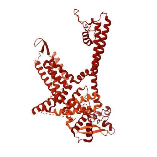
8DRP

Focus/local refined map in C4 of signal subtracted RyR1 particles


Experimental Data Snapshot

  • Method: ELECTRON MICROSCOPY
  • Resolution: 2.84 Å
  • Aggregation State: PARTICLE 
  • Reconstruction Method: SINGLE PARTICLE 

wwPDB Validation   3D Report Full Report


This is version 1.2 of the entry. See complete history


Literature

Cryo-EM analysis of scorpion toxin binding to Ryanodine Receptors reveals subconductance that is abolished by PKA phosphorylation.

Haji-Ghassemi, O.Chen, Y.S.Woll, K.Gurrola, G.B.Valdivia, C.R.Cai, W.Li, S.Valdivia, H.H.Van Petegem, F.

(2023) Sci Adv 9: eadf4936-eadf4936

  • DOI: https://doi.org/10.1126/sciadv.adf4936
  • Primary Citation of Related Structures:  
    8DRP, 8DTB, 8DUJ, 8DVE

  • PubMed Abstract: 

    Calcins are peptides from scorpion venom with the unique ability to cross cell membranes, gaining access to intracellular targets. Ryanodine Receptors (RyR) are intracellular ion channels that control release of Ca 2+ from the endoplasmic and sarcoplasmic reticulum. Calcins target RyRs and induce long-lived subconductance states, whereby single-channel currents are decreased. We used cryo-electron microscopy to reveal the binding and structural effects of imperacalcin, showing that it opens the channel pore and causes large asymmetry throughout the cytosolic assembly of the tetrameric RyR. This also creates multiple extended ion conduction pathways beyond the transmembrane region, resulting in subconductance. Phosphorylation of imperacalcin by protein kinase A prevents its binding to RyR through direct steric hindrance, showing how posttranslational modifications made by the host organism can determine the fate of a natural toxin. The structure provides a direct template for developing calcin analogs that result in full channel block, with potential to treat RyR-related disorders.


  • Organizational Affiliation

    Department of Biochemistry and Molecular Biology, Life Sciences Centre, University of British Columbia, Vancouver, BC, Canada.


Macromolecules
Find similar proteins by:  (by identity cutoff)  |  3D Structure
Entity ID: 1
MoleculeChains Sequence LengthOrganismDetailsImage
Ryanodine receptor 1
A, B, C, D
5,037Oryctolagus cuniculusMutation(s): 0 
Membrane Entity: Yes 
UniProt
Find proteins for P11716 (Oryctolagus cuniculus)
Explore P11716 
Go to UniProtKB:  P11716
Entity Groups  
Sequence Clusters30% Identity50% Identity70% Identity90% Identity95% Identity100% Identity
UniProt GroupP11716
Sequence Annotations
Expand
  • Reference Sequence
Small Molecules
Ligands 3 Unique
IDChains Name / Formula / InChI Key2D Diagram3D Interactions
ATP
Query on ATP

Download Ideal Coordinates CCD File 
F [auth A],
I [auth B],
L [auth C],
O [auth D]
ADENOSINE-5'-TRIPHOSPHATE
C10 H16 N5 O13 P3
ZKHQWZAMYRWXGA-KQYNXXCUSA-N
CFF
Query on CFF

Download Ideal Coordinates CCD File 
E [auth A],
H [auth B],
K [auth C],
N [auth D]
CAFFEINE
C8 H10 N4 O2
RYYVLZVUVIJVGH-UHFFFAOYSA-N
ZN
Query on ZN

Download Ideal Coordinates CCD File 
G [auth A],
J [auth B],
M [auth C],
P [auth D]
ZINC ION
Zn
PTFCDOFLOPIGGS-UHFFFAOYSA-N
Experimental Data & Validation

Experimental Data

  • Method: ELECTRON MICROSCOPY
  • Resolution: 2.84 Å
  • Aggregation State: PARTICLE 
  • Reconstruction Method: SINGLE PARTICLE 
EM Software:
TaskSoftware PackageVersion
RECONSTRUCTIONcryoSPARC3.2

Structure Validation

View Full Validation Report


Entry History & Funding Information

Deposition Data


Funding OrganizationLocationGrant Number
Canadian Institutes of Health Research (CIHR)CanadaPJT-159601
Canadian Institutes of Health Research (CIHR)CanadaMFE-381863

Revision History  (Full details and data files)

  • Version 1.0: 2023-05-31
    Type: Initial release
  • Version 1.1: 2023-06-14
    Changes: Database references
  • Version 1.2: 2024-03-13
    Changes: Data collection, Source and taxonomy