8E0A

Crystal structure of human Sar1b

  • Classification: SIGNALING PROTEIN
  • Organism(s): Homo sapiens
  • Expression System: Escherichia coli
  • Mutation(s): No 

  • Deposited: 2022-08-08 Released: 2022-11-30 
  • Deposition Author(s): Huang, Q.
  • Funding Organization(s): National Institutes of Health/National Institute of General Medical Sciences (NIH/NIGMS)

Experimental Data Snapshot

  • Method: X-RAY DIFFRACTION
  • Resolution: 1.80 Å
  • R-Value Free: 0.209 
  • R-Value Work: 0.169 
  • R-Value Observed: 0.171 

wwPDB Validation   3D Report Full Report


Ligand Structure Quality Assessment 


This is version 1.2 of the entry. See complete history


Literature

The alarmone ppGpp selectively inhibits the isoform A of the human small GTPase Sar1.

Huang, Q.Szebenyi, D.M.E.

(2023) Proteins 91: 518-531

  • DOI: https://doi.org/10.1002/prot.26445
  • Primary Citation of Related Structures:  
    8DZM, 8DZN, 8DZO, 8DZT, 8E0A, 8E0B, 8E0C, 8E0D, 8E0H

  • PubMed Abstract: 

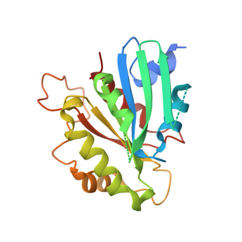
    Transport of newly synthesized proteins from endoplasmic reticulum (ER) to Golgi is mediated by coat protein complex II (COPII). The assembly and disassembly of COPII vesicles is regulated by the molecular switch Sar1, which is a small GTPase and a component of COPII. Usually a small GTPase binds GDP (inactive form) or GTP (active form). Mammals have two Sar1 isoforms, Sar1a and Sar1b, that have approximately 90% sequence identity. Some experiments demonstrated that these two isoforms had distinct but overlapping functions. Here we found another instance of differing behavior: the alarmone ppGpp could bind to and inhibit the GTPase activity of human Sar1a but could not inhibit the GTPase activity of human Sar1b. The crystal structures of Sar1a⋅ppGpp and Sar1b⋅GDP have been determined. Superposition of the structures shows that ppGpp binds to the nucleotide-binding pocket, its guanosine base, ribose ring and 5'-diphosphate occupying nearly the same positions as for GDP. However, its 3'-diphosphate points away from the active site and, hence, away from the surface of the protein. The overall structure of Sar1a⋅ppGpp is more similar to Sar1b⋅GDP than to Sar1b⋅GTP. We also find that the Asp140-Arg138-water-ligand interaction net is important for the binding of ppGpp to Sar1a. This study provides further evidence showing that there are biochemical differences between the Sar1a and Sar1b isoforms of Sar1.


  • Organizational Affiliation

    Cornell High Energy Synchrotron Source (CHESS), Cornell University, Ithaca, New York, USA.


Macromolecules
Find similar proteins by:  (by identity cutoff)  |  3D Structure
Entity ID: 1
MoleculeChains Sequence LengthOrganismDetailsImage
GTP-binding protein SAR1b
A, B
198Homo sapiensMutation(s): 0 
Gene Names: SAR1BSARA2SARB
UniProt & NIH Common Fund Data Resources
Find proteins for Q9Y6B6 (Homo sapiens)
Explore Q9Y6B6 
Go to UniProtKB:  Q9Y6B6
PHAROS:  Q9Y6B6
GTEx:  ENSG00000152700 
Entity Groups  
Sequence Clusters30% Identity50% Identity70% Identity90% Identity95% Identity100% Identity
UniProt GroupQ9Y6B6
Sequence Annotations
Expand
  • Reference Sequence
Small Molecules
Ligands 2 Unique
IDChains Name / Formula / InChI Key2D Diagram3D Interactions
GDP (Subject of Investigation/LOI)
Query on GDP

Download Ideal Coordinates CCD File 
G [auth A],
K [auth B]
GUANOSINE-5'-DIPHOSPHATE
C10 H15 N5 O11 P2
QGWNDRXFNXRZMB-UUOKFMHZSA-N
SO4 (Subject of Investigation/LOI)
Query on SO4

Download Ideal Coordinates CCD File 
C [auth A]
D [auth A]
E [auth A]
F [auth A]
H [auth B]
C [auth A],
D [auth A],
E [auth A],
F [auth A],
H [auth B],
I [auth B],
J [auth B]
SULFATE ION
O4 S
QAOWNCQODCNURD-UHFFFAOYSA-L
Experimental Data & Validation

Experimental Data

  • Method: X-RAY DIFFRACTION
  • Resolution: 1.80 Å
  • R-Value Free: 0.209 
  • R-Value Work: 0.169 
  • R-Value Observed: 0.171 
  • Space Group: P 1 21 1
Unit Cell:
Length ( Å )Angle ( ˚ )
a = 52.966α = 90
b = 61.485β = 107.34
c = 70.589γ = 90
Software Package:
Software NamePurpose
PHENIXrefinement
PDB_EXTRACTdata extraction
HKL-2000data reduction
HKL-2000data scaling
PHENIXphasing

Structure Validation

View Full Validation Report


Ligand Structure Quality Assessment 


Entry History & Funding Information

Deposition Data

  • Released Date: 2022-11-30 
  • Deposition Author(s): Huang, Q.

Funding OrganizationLocationGrant Number
National Institutes of Health/National Institute of General Medical Sciences (NIH/NIGMS)United StatesR01 GM040654
National Institutes of Health/National Institute of General Medical Sciences (NIH/NIGMS)United StatesR01 CA163255

Revision History  (Full details and data files)

  • Version 1.0: 2022-11-30
    Type: Initial release
  • Version 1.1: 2023-03-22
    Changes: Database references
  • Version 1.2: 2023-10-25
    Changes: Data collection, Refinement description