Download Mendeley
Comprehensive solution NMR characterization of bivalirudin, a direct thrombin inhibitor peptide, and its process impurities by heteronuclear spectral fingerprinting at natural isotopic abundance.
Mishra, S.H., Kaur, A., Vu, D., Bhavaraju, S.(2026) J Pharm Biomed Anal 281: 117684-117684
- PubMed: 42542115 Search on PubMed
- DOI: https://doi.org/10.1016/j.jpba.2026.117684
- Primary Citation Related Structures:
8EF4 - PubMed Abstract:
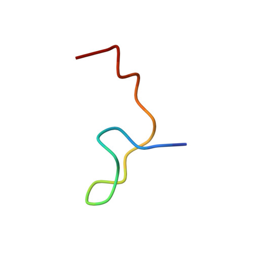
Bivalirudin is a 20-residue synthetic peptide that functions as a bivalent thrombin inhibitor by simultaneously engaging thrombin's active site and exosite I. Despite its widespread clinical use, comprehensive NMR-based characterization of bivalirudin and its process-related impurities has not been reported. Here, we present the first complete heteronuclear NMR analysis of bivalirudin and two major impurities-[Asp9]-bivalirudin (a deamidation-related impurity standard) and [desGlu13]-bivalirudin (a synthesis deletion)-at natural isotopic abundance. Using a combination of 1D and 2D 1 H, 13 C, and 15 N NMR experiments in DMSO-d₆ and aqueous solution, we achieved near-complete backbone and side-chain assignments, now deposited in the BMRB. We demonstrate that 2D heteronuclear amide maps can unambiguously differentiate the parent peptide from its process impurities. Additionally, we report the first solution-state NMR structure of free bivalirudin, revealing a compact architecture wherein the active site- and exosite-binding segments are spatially proximate, potentially reflecting a pre-binding intermediate. This study establishes a practical spectral reference framework and simplified 2D heteronuclear spectral fingerprints for structural integrity assessment and impurity tracking of bivalirudin.
- National Institute of Standards and Technology, (formerly at US Pharmacopeial Convention, Rockville, MD, USA), Gaithersburg, MD, USA; Institute for Biosciences and Biotechnology Research, University of Maryland, College Park, Rockville, MD, USA. Electronic address: shmishra@umd.edu.
Organizational Affiliation: