8ENT

Interleukin-21 signaling complex with IL-21R and IL-2Rg


Experimental Data Snapshot

  • Method: X-RAY DIFFRACTION
  • Resolution: 2.83 Å
  • R-Value Free: 
    0.300 (Depositor), 0.300 (DCC) 
  • R-Value Work: 
    0.254 (Depositor), 0.250 (DCC) 
  • R-Value Observed: 
    0.255 (Depositor) 

Starting Models: experimental
View more details

wwPDB Validation   3D Report Full Report


Ligand Structure Quality Assessment 

Created with Raphaël 2.3.0Worse 01 BetterLigand structure goodness of fit to experimental dataBest fitted NAGClick on this verticalbar to view details

This is version 1.2 of the entry. See complete history


Literature

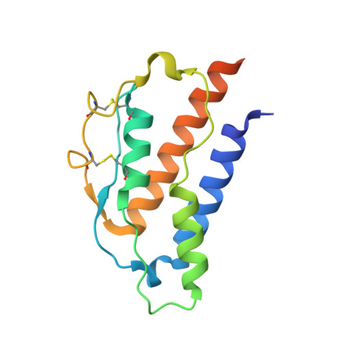
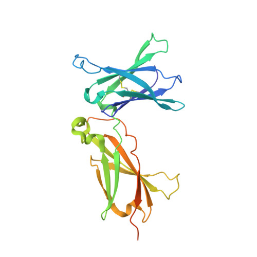
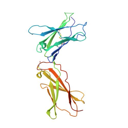
A structural blueprint for interleukin-21 signal modulation.

Abhiraman, G.C.Bruun, T.U.J.Caveney, N.A.Su, L.L.Saxton, R.A.Yin, Q.Tang, S.Davis, M.M.Jude, K.M.Garcia, K.C.

(2023) Cell Rep 42: 112657-112657

  • DOI: https://doi.org/10.1016/j.celrep.2023.112657
  • Primary Citation of Related Structures:  
    8ENT

  • PubMed Abstract: 

    Interleukin-21 (IL-21) plays a critical role in generating immunological memory by promoting the germinal center reaction, yet clinical use of IL-21 remains challenging because of its pleiotropy and association with autoimmune disease. To better understand the structural basis of IL-21 signaling, we determine the structure of the IL-21-IL-21R-γc ternary signaling complex by X-ray crystallography and a structure of a dimer of trimeric complexes using cryo-electron microscopy. Guided by the structure, we design analogs of IL-21 by introducing substitutions to the IL-21-γc interface. These IL-21 analogs act as partial agonists that modulate downstream activation of pS6, pSTAT3, and pSTAT1. These analogs exhibit differential activity on T and B cell subsets and modulate antibody production in human tonsil organoids. These results clarify the structural basis of IL-21 signaling and offer a potential strategy for tunable manipulation of humoral immunity.


  • Organizational Affiliation

    Department of Molecular and Cellular Physiology, Stanford University School of Medicine, 279 Campus Drive, Stanford, CA 94305, USA; Program in Immunology, Stanford University School of Medicine, Stanford, CA 94305, USA.


Macromolecules
Find similar proteins by:  (by identity cutoff)  |  3D Structure
Entity ID: 1
MoleculeChains Sequence LengthOrganismDetailsImage
Interleukin-21
A, D, G, J
144Homo sapiensMutation(s): 1 
Gene Names: IL21
UniProt & NIH Common Fund Data Resources
Find proteins for Q9HBE4 (Homo sapiens)
Explore Q9HBE4 
Go to UniProtKB:  Q9HBE4
PHAROS:  Q9HBE4
GTEx:  ENSG00000138684 
Entity Groups  
Sequence Clusters30% Identity50% Identity70% Identity90% Identity95% Identity100% Identity
UniProt GroupQ9HBE4
Sequence Annotations
Expand
  • Reference Sequence
Find similar proteins by:  (by identity cutoff)  |  3D Structure
Entity ID: 2
MoleculeChains Sequence LengthOrganismDetailsImage
Interleukin-21 receptor
B, E, H, K
237Homo sapiensMutation(s): 4 
Gene Names: IL21RNILRUNQ3121/PRO10273
UniProt & NIH Common Fund Data Resources
Find proteins for Q9HBE5 (Homo sapiens)
Explore Q9HBE5 
Go to UniProtKB:  Q9HBE5
PHAROS:  Q9HBE5
GTEx:  ENSG00000103522 
Entity Groups  
Sequence Clusters30% Identity50% Identity70% Identity90% Identity95% Identity100% Identity
UniProt GroupQ9HBE5
Glycosylation
Glycosylation Sites: 1Go to GlyGen: Q9HBE5-1
Sequence Annotations
Expand
  • Reference Sequence
Find similar proteins by:  (by identity cutoff)  |  3D Structure
Entity ID: 3
MoleculeChains Sequence LengthOrganismDetailsImage
Cytokine receptor common subunit gamma
C, F, I
207Homo sapiensMutation(s): 1 
Gene Names: IL2RG
UniProt & NIH Common Fund Data Resources
Find proteins for P31785 (Homo sapiens)
Explore P31785 
Go to UniProtKB:  P31785
PHAROS:  P31785
GTEx:  ENSG00000147168 
Entity Groups  
Sequence Clusters30% Identity50% Identity70% Identity90% Identity95% Identity100% Identity
UniProt GroupP31785
Glycosylation
Glycosylation Sites: 3Go to GlyGen: P31785-1
Sequence Annotations
Expand
  • Reference Sequence
Oligosaccharides

Help

Entity ID: 4
MoleculeChains Length2D Diagram Glycosylation3D Interactions
2-acetamido-2-deoxy-beta-D-glucopyranose-(1-4)-2-acetamido-2-deoxy-beta-D-glucopyranose
L, M, O
2N-Glycosylation
Glycosylation Resources
GlyTouCan:  G42666HT
GlyCosmos:  G42666HT
GlyGen:  G42666HT
Entity ID: 5
MoleculeChains Length2D Diagram Glycosylation3D Interactions
beta-D-mannopyranose-(1-4)-2-acetamido-2-deoxy-beta-D-glucopyranose-(1-4)-2-acetamido-2-deoxy-beta-D-glucopyranose
N
3N-Glycosylation
Glycosylation Resources
GlyTouCan:  G15407YE
GlyCosmos:  G15407YE
GlyGen:  G15407YE
Small Molecules
Ligands 2 Unique
IDChains Name / Formula / InChI Key2D Diagram3D Interactions
NAG
Query on NAG

Download Ideal Coordinates CCD File 
AA [auth I]
BA [auth I]
P [auth C]
R [auth C]
U [auth E]
2-acetamido-2-deoxy-beta-D-glucopyranose
C8 H15 N O6
OVRNDRQMDRJTHS-FMDGEEDCSA-N
EDO
Query on EDO

Download Ideal Coordinates CCD File 
CA [auth I]
DA [auth I]
EA [auth K]
FA [auth K]
GA [auth K]
1,2-ETHANEDIOL
C2 H6 O2
LYCAIKOWRPUZTN-UHFFFAOYSA-N
Experimental Data & Validation

Experimental Data

  • Method: X-RAY DIFFRACTION
  • Resolution: 2.83 Å
  • R-Value Free:  0.300 (Depositor), 0.300 (DCC) 
  • R-Value Work:  0.254 (Depositor), 0.250 (DCC) 
  • R-Value Observed: 0.255 (Depositor) 
Space Group: P 1
Unit Cell:
Length ( Å )Angle ( ˚ )
a = 66.167α = 83.34
b = 66.464β = 83.71
c = 162.684γ = 73.1
Software Package:
Software NamePurpose
PHENIXrefinement
PHENIXmodel building
XDSdata scaling
XDSdata reduction
PHENIXphasing

Structure Validation

View Full Validation Report


Ligand Structure Quality Assessment 

Created with Raphaël 2.3.0Worse 01 BetterLigand structure goodness of fit to experimental dataBest fitted NAGClick on this verticalbar to view details

Entry History & Funding Information

Deposition Data


Funding OrganizationLocationGrant Number
National Institutes of Health/National Institute Of Allergy and Infectious Diseases (NIH/NIAID)United StatesRO1-AI51321
Bill & Melinda Gates FoundationUnited States--

Revision History  (Full details and data files)

  • Version 1.0: 2023-06-28
    Type: Initial release
  • Version 1.1: 2023-10-25
    Changes: Data collection, Refinement description
  • Version 1.2: 2024-11-20
    Changes: Structure summary