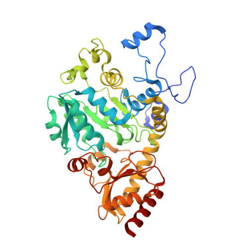
8ERB

Crystal structure of Fub7 in complex with vinylglycine ketimine


Experimental Data Snapshot

  • Method: X-RAY DIFFRACTION
  • Resolution: 1.98 Å
  • R-Value Free: 0.226 
  • R-Value Work: 0.200 
  • R-Value Observed: 0.202 

wwPDB Validation   3D Report Full Report


Ligand Structure Quality Assessment 


This is version 1.2 of the entry. See complete history


Literature

Molecular and Structural Basis for C gamma-C Bond Formation by PLP-Dependent Enzyme Fub7.

Liu, S.Yeh, C.Reavill, C.Jones, B.Zou, Y.Hai, Y.

(2024) Angew Chem Int Ed Engl 63: e202317161-e202317161

  • DOI: https://doi.org/10.1002/anie.202317161
  • Primary Citation of Related Structures:  
    8EQW, 8ERB, 8ERJ

  • PubMed Abstract: 

    Pyridoxal 5'-phosphate (PLP)-dependent enzymes that catalyze γ-replacement reactions are prevalent, yet their utilization of carbon nucleophile substrates is rare. The recent discovery of two PLP-dependent enzymes, CndF and Fub7, has unveiled unique C-C bond forming capabilities, enabling the biocatalytic synthesis of alkyl- substituted pipecolic acids from O-acetyl-L-homoserine and β-keto acid or aldehyde derived enolates. This breakthrough presents fresh avenues for the biosynthesis of pipecolic acid derivatives. However, the catalytic mechanisms of these enzymes remain elusive, and a dearth of structural information hampers their extensive application. Here, we have broadened the catalytic scope of Fub7 by employing ketone-derived enolates as carbon nucleophiles, revealing Fub7's capacity for substrate-dependent regioselective α-alkylation of unsymmetrical ketones. Through an integrated approach combining X-ray crystallography, spectroscopy, mutagenesis, and computational docking studies, we offer a detailed mechanistic insight into Fub7 catalysis. Our findings elucidate the structural basis for its substrate specificity, stereoselectivity, and regioselectivity. Our work sets the stage ready for subsequent protein engineering effort aimed at expanding the synthetic utility of Fub7, potentially unlocking novel methods to access a broader array of noncanonical amino acids.


  • Organizational Affiliation

    Department of Chemistry and Biochemistry, University of California Santa Barbara, 93110, Santa Barbara, CA, USA.


Macromolecules
Find similar proteins by:  (by identity cutoff)  |  3D Structure
Entity ID: 1
MoleculeChains Sequence LengthOrganismDetailsImage
Sulfhydrylase FUB7433Fusarium fujikuroiMutation(s): 0 
Gene Names: FUB7
EC: 2.5.1
UniProt
Find proteins for S0DUX5 (Gibberella fujikuroi (strain CBS 195.34 / IMI 58289 / NRRL A-6831))
Explore S0DUX5 
Go to UniProtKB:  S0DUX5
Entity Groups  
Sequence Clusters30% Identity50% Identity70% Identity90% Identity95% Identity100% Identity
UniProt GroupS0DUX5
Sequence Annotations
Expand
  • Reference Sequence
Small Molecules
Ligands 1 Unique
IDChains Name / Formula / InChI Key2D Diagram3D Interactions
WBJ (Subject of Investigation/LOI)
Query on WBJ

Download Ideal Coordinates CCD File 
AA [auth M]
BA [auth Q]
CA [auth N]
DA [auth E]
EA [auth L]
AA [auth M],
BA [auth Q],
CA [auth N],
DA [auth E],
EA [auth L],
FA [auth K],
Q [auth A],
R [auth B],
S [auth C],
T [auth D],
U [auth G],
V [auth H],
W [auth F],
X [auth I],
Y [auth P],
Z [auth J]
(2E)-2-[({3-hydroxy-2-methyl-5-[(phosphonooxy)methyl]pyridin-4-yl}methyl)imino]but-3-enoic acid
C12 H15 N2 O7 P
ILCWREDNKMRIGC-GXDHUFHOSA-N
Experimental Data & Validation

Experimental Data

  • Method: X-RAY DIFFRACTION
  • Resolution: 1.98 Å
  • R-Value Free: 0.226 
  • R-Value Work: 0.200 
  • R-Value Observed: 0.202 
  • Space Group: P 1 21 1
Unit Cell:
Length ( Å )Angle ( ˚ )
a = 146.703α = 90
b = 193.364β = 99.6
c = 149.913γ = 90
Software Package:
Software NamePurpose
PHENIXrefinement
HKL-3000data reduction
HKL-3000data scaling
PHASERphasing

Structure Validation

View Full Validation Report


Ligand Structure Quality Assessment 


Entry History & Funding Information

Deposition Data

  • Released Date: 2023-10-18 
  • Deposition Author(s): Hai, Y.

Funding OrganizationLocationGrant Number
Other governmentUnited States--

Revision History  (Full details and data files)

  • Version 1.0: 2023-10-18
    Type: Initial release
  • Version 1.1: 2024-02-14
    Changes: Database references
  • Version 1.2: 2024-03-27
    Changes: Database references