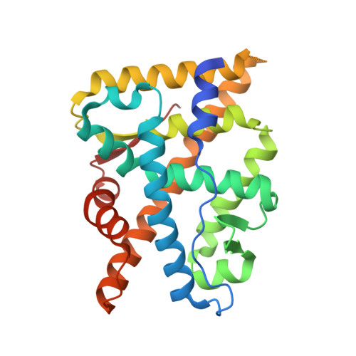
8FGY

Crystal structure of mutant Androgen Receptor ligand binding domain L702H/H875Y/F877L/T878A with DHT


Experimental Data Snapshot

  • Method: X-RAY DIFFRACTION
  • Resolution: 2.20 Å
  • R-Value Free: 0.246 
  • R-Value Work: 0.195 
  • R-Value Observed: 0.200 

Starting Model: experimental
View more details

wwPDB Validation   3D Report Full Report


Ligand Structure Quality Assessment 


This is version 1.1 of the entry. See complete history


Literature

A partially open conformation of an androgen receptor ligand-binding domain with drug-resistance mutations.

Doamekpor, S.K.Peng, P.Xu, R.Ma, L.Tong, Y.Tong, L.

(2023) Acta Crystallogr F Struct Biol Commun 79: 95-104

  • DOI: https://doi.org/10.1107/S2053230X23002224
  • Primary Citation of Related Structures:  
    8FGY, 8FGZ, 8FH0, 8FH1, 8FH2

  • PubMed Abstract: 

    Mutations in the androgen receptor (AR) ligand-binding domain (LBD) can cause resistance to drugs used to treat prostate cancer. Commonly found mutations include L702H, W742C, H875Y, F877L and T878A, while the F877L mutation can convert second-generation antagonists such as enzalutamide and apalutamide into agonists. However, pruxelutamide, another second-generation AR antagonist, has no agonist activity with the F877L and F877L/T878A mutants and instead maintains its inhibitory activity against them. Here, it is shown that the quadruple mutation L702H/H875Y/F877L/T878A increases the soluble expression of AR LBD in complex with pruxelutamide in Escherichia coli. The crystal structure of the quadruple mutant in complex with the agonist dihydrotestosterone (DHT) reveals a partially open conformation of the AR LBD due to conformational changes in the loop connecting helices H11 and H12 (the H11-H12 loop) and Leu881. This partially open conformation creates a larger ligand-binding site for AR. Additional structural studies suggest that both the L702H and F877L mutations are important for conformational changes. This structural variability in the AR LBD could affect ligand binding as well as the resistance to antagonists.


  • Organizational Affiliation

    Department of Biological Sciences, Columbia University, New York, NY 10027, USA.


Macromolecules
Find similar proteins by:  (by identity cutoff)  |  3D Structure
Entity ID: 1
MoleculeChains Sequence LengthOrganismDetailsImage
Androgen receptor258Homo sapiensMutation(s): 4 
Gene Names: ARDHTRNR3C4
UniProt & NIH Common Fund Data Resources
Find proteins for P10275 (Homo sapiens)
Explore P10275 
Go to UniProtKB:  P10275
PHAROS:  P10275
GTEx:  ENSG00000169083 
Entity Groups  
Sequence Clusters30% Identity50% Identity70% Identity90% Identity95% Identity100% Identity
UniProt GroupP10275
Sequence Annotations
Expand
  • Reference Sequence
Small Molecules
Ligands 1 Unique
IDChains Name / Formula / InChI Key2D Diagram3D Interactions
DHT (Subject of Investigation/LOI)
Query on DHT

Download Ideal Coordinates CCD File 
B [auth A]5-ALPHA-DIHYDROTESTOSTERONE
C19 H30 O2
NVKAWKQGWWIWPM-ABEVXSGRSA-N
Binding Affinity Annotations 
IDSourceBinding Affinity
DHT BindingDB:  8FGY Ki: min: 0.2, max: 10 (nM) from 12 assay(s)
Kd: 0.25 (nM) from 1 assay(s)
IC50: min: 0.05, max: 18.5 (nM) from 14 assay(s)
EC50: min: 0.05, max: 20 (nM) from 16 assay(s)
Experimental Data & Validation

Experimental Data

  • Method: X-RAY DIFFRACTION
  • Resolution: 2.20 Å
  • R-Value Free: 0.246 
  • R-Value Work: 0.195 
  • R-Value Observed: 0.200 
  • Space Group: P 21 21 21
Unit Cell:
Length ( Å )Angle ( ˚ )
a = 56.413α = 90
b = 66.096β = 90
c = 73.382γ = 90
Software Package:
Software NamePurpose
PHENIXrefinement
XDSdata scaling
XDSdata reduction
PHENIXphasing

Structure Validation

View Full Validation Report


Ligand Structure Quality Assessment 


Entry History & Funding Information

Deposition Data


Funding OrganizationLocationGrant Number
Other private--

Revision History  (Full details and data files)

  • Version 1.0: 2023-04-12
    Type: Initial release
  • Version 1.1: 2024-05-22
    Changes: Data collection