8FLL

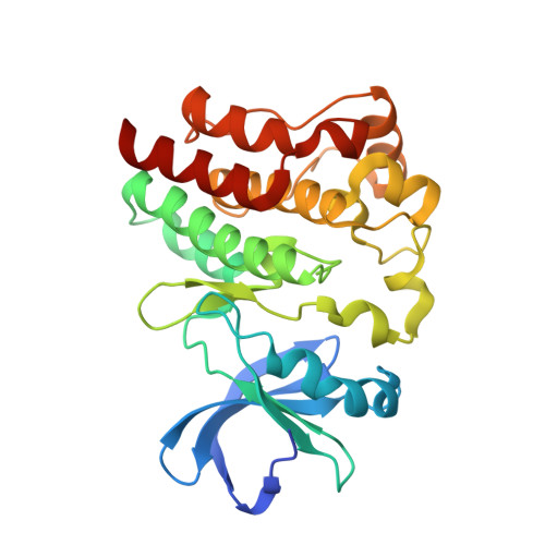
Crystal structure of BTK kinase domain in complex with pirtobrutinib


Experimental Data Snapshot

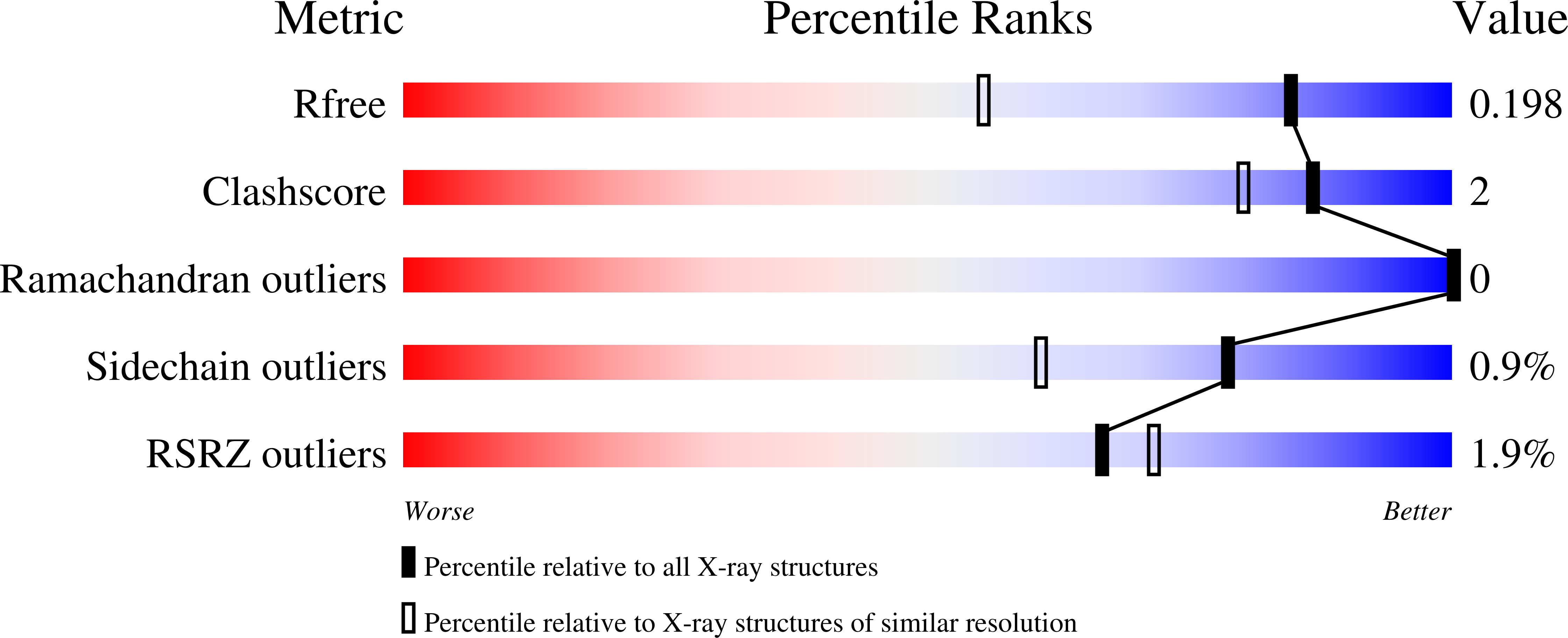
  • Method: X-RAY DIFFRACTION
  • Resolution: 1.50 Å
  • R-Value Free: 0.202 
  • R-Value Work: 0.185 
  • R-Value Observed: 0.186 

Starting Model: experimental
View more details

wwPDB Validation   3D Report Full Report


Ligand Structure Quality Assessment 


This is version 1.4 of the entry. See complete history


Literature

Preclinical characterization of pirtobrutinib, a highly selective, noncovalent (reversible) BTK inhibitor.

Gomez, E.B.Ebata, K.Randeria, H.S.Rosendahl, M.S.Cedervall, E.P.Morales, T.H.Hanson, L.M.Brown, N.E.Gong, X.Stephens, J.Wu, W.Lippincott, I.Ku, K.S.Walgren, R.A.Abada, P.B.Ballard, J.A.Allerston, C.K.Brandhuber, B.J.

(2023) Blood 142: 62-72

  • DOI: https://doi.org/10.1182/blood.2022018674
  • Primary Citation of Related Structures:  
    8FLL, 8FLN

  • PubMed Abstract: 

    Bruton tyrosine kinase (BTK), a nonreceptor tyrosine kinase, is a major therapeutic target for B-cell-driven malignancies. However, approved covalent BTK inhibitors (cBTKis) are associated with treatment limitations because of off-target side effects, suboptimal oral pharmacology, and development of resistance mutations (eg, C481) that prevent inhibitor binding. Here, we describe the preclinical profile of pirtobrutinib, a potent, highly selective, noncovalent (reversible) BTK inhibitor. Pirtobrutinib binds BTK with an extensive network of interactions to BTK and water molecules in the adenosine triphosphate binding region and shows no direct interaction with C481. Consequently, pirtobrutinib inhibits both BTK and BTK C481 substitution mutants in enzymatic and cell-based assays with similar potencies. In differential scanning fluorimetry studies, BTK bound to pirtobrutinib exhibited a higher melting temperature than cBTKi-bound BTK. Pirtobrutinib, but not cBTKis, prevented Y551 phosphorylation in the activation loop. These data suggest that pirtobrutinib uniquely stabilizes BTK in a closed, inactive conformation. Pirtobrutinib inhibits BTK signaling and cell proliferation in multiple B-cell lymphoma cell lines, and significantly inhibits tumor growth in human lymphoma xenografts in vivo. Enzymatic profiling showed that pirtobrutinib was highly selective for BTK in >98% of the human kinome, and in follow-up cellular studies pirtobrutinib retained >100-fold selectivity over other tested kinases. Collectively, these findings suggest that pirtobrutinib represents a novel BTK inhibitor with improved selectivity and unique pharmacologic, biophysical, and structural attributes with the potential to treat B-cell-driven cancers with improved precision and tolerability. Pirtobrutinib is being tested in phase 3 clinical studies for a variety of B-cell malignancies.


  • Organizational Affiliation

    Loxo@Lilly, Louisville, CO.


Macromolecules
Find similar proteins by:  (by identity cutoff)  |  3D Structure
Entity ID: 1
MoleculeChains Sequence LengthOrganismDetailsImage
Tyrosine-protein kinase BTK273Homo sapiensMutation(s): 0 
Gene Names: BTKAGMX1ATKBPK
EC: 2.7.10.2
UniProt & NIH Common Fund Data Resources
Find proteins for Q06187 (Homo sapiens)
Explore Q06187 
Go to UniProtKB:  Q06187
PHAROS:  Q06187
GTEx:  ENSG00000010671 
Entity Groups  
Sequence Clusters30% Identity50% Identity70% Identity90% Identity95% Identity100% Identity
UniProt GroupQ06187
Sequence Annotations
Expand
  • Reference Sequence
Small Molecules
Ligands 4 Unique
IDChains Name / Formula / InChI Key2D Diagram3D Interactions
Y7W (Subject of Investigation/LOI)
Query on Y7W

Download Ideal Coordinates CCD File 
B [auth A]pirtobrutinib
C22 H21 F4 N5 O3
FWZAWAUZXYCBKZ-NSHDSACASA-N
BTB
Query on BTB

Download Ideal Coordinates CCD File 
F [auth A]2-[BIS-(2-HYDROXY-ETHYL)-AMINO]-2-HYDROXYMETHYL-PROPANE-1,3-DIOL
C8 H19 N O5
OWMVSZAMULFTJU-UHFFFAOYSA-N
SO4
Query on SO4

Download Ideal Coordinates CCD File 
C [auth A]SULFATE ION
O4 S
QAOWNCQODCNURD-UHFFFAOYSA-L
GOL
Query on GOL

Download Ideal Coordinates CCD File 
D [auth A],
E [auth A]
GLYCEROL
C3 H8 O3
PEDCQBHIVMGVHV-UHFFFAOYSA-N
Binding Affinity Annotations 
IDSourceBinding Affinity
Y7W BindingDB:  8FLL IC50: 1 (nM) from 1 assay(s)
Experimental Data & Validation

Experimental Data

  • Method: X-RAY DIFFRACTION
  • Resolution: 1.50 Å
  • R-Value Free: 0.202 
  • R-Value Work: 0.185 
  • R-Value Observed: 0.186 
  • Space Group: P 21 21 21
Unit Cell:
Length ( Å )Angle ( ˚ )
a = 41.482α = 90
b = 67.269β = 90
c = 90.383γ = 90
Software Package:
Software NamePurpose
BUSTERrefinement
autoPROCdata reduction
autoPROCdata scaling
PHASERphasing

Structure Validation

View Full Validation Report


Ligand Structure Quality Assessment 


Entry History & Funding Information

Deposition Data


Funding OrganizationLocationGrant Number
Not funded--

Revision History  (Full details and data files)

  • Version 1.0: 2023-03-01
    Type: Initial release
  • Version 1.1: 2023-03-08
    Changes: Refinement description, Structure summary
  • Version 1.2: 2023-03-15
    Changes: Structure summary
  • Version 1.3: 2023-07-12
    Changes: Database references
  • Version 1.4: 2024-05-22
    Changes: Data collection