8FUJ | pdb_00008fuj

HIV-1 wild type protease with GRL-03419A, with N-(2,5-dimethylphenyl)-4-(pyridin-3-yl)pyrimidin-2-amine as P2-P3 group and 3,5-difluorophenylmethyl as the P1 group


Experimental Data Snapshot

  • Method: X-RAY DIFFRACTION
  • Resolution: 1.12 Å
  • R-Value Free: 
    0.136 (Depositor), 0.135 (DCC) 
  • R-Value Work: 
    0.121 (Depositor), 0.120 (DCC) 
  • R-Value Observed: 
    0.121 (Depositor) 

Starting Model: experimental
View more details

wwPDB Validation 3D Report Full Report

Validation slider image for 8FUJ

Ligand Structure Quality Assessment 


This is version 1.1 of the entry. See complete history

Literature

Exploration of imatinib and nilotinib-derived templates as the P2-Ligand for HIV-1 protease inhibitors: Design, synthesis, protein X-ray structural studies, and biological evaluation.

Ghosh, A.K.Mishevich, J.L.Kovela, S.Shaktah, R.Ghosh, A.K.Johnson, M.Wang, Y.F.Wong-Sam, A.Agniswamy, J.Amano, M.Takamatsu, Y.Hattori, S.I.Weber, I.T.Mitsuya, H.

(2023) Eur J Med Chem 255: 115385-115385

  • DOI: https://doi.org/10.1016/j.ejmech.2023.115385
  • Primary Citation Related Structures: 
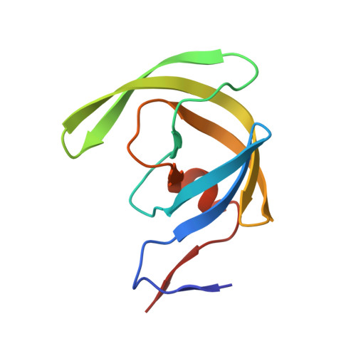
    8FUI, 8FUJ

  • PubMed Abstract: 

    Structure-based design, synthesis, X-ray structural studies, and biological evaluation of a new series of potent HIV-1 protease inhibitors are described. These inhibitors contain various pyridyl-pyrimidine, aryl thiazole or alkylthiazole derivatives as the P2 ligands in combination with darunavir-like hydroxyethylamine sulfonamide isosteres. These heterocyclic ligands are inherent to kinase inhibitor drugs, such as nilotinib and imatinib. These ligands are designed to make hydrogen bonding interactions with the backbone atoms in the S2 subsite of HIV-1 protease. Various benzoic acid derivatives have been synthesized and incorporation of these ligands provided potent inhibitors that exhibited subnanomolar level protease inhibitory activity and low nanomolar level antiviral activity. Two high resolution X-ray structures of inhibitor-bound HIV-1 protease were determined. These structures provided important ligand-binding site interactions for further optimization of this class of protease inhibitors.


  • Organizational Affiliation
    • Department of Chemistry and Department of Medicinal Chemistry, Purdue University, West Lafayette, IN, 47907, United States. Electronic address: akghosh@purdue.edu.

Macromolecule Content 

  • Total Structure Weight: 22.56 kDa 
  • Atom Count: 2,255 
  • Modeled Residue Count: 198 
  • Deposited Residue Count: 198 
  • Unique protein chains: 1

Macromolecules

Find similar proteins by:|  3D Structure
Entity ID: 1
MoleculeChains  Sequence LengthOrganismDetailsImage
Protease
A, B
99Human immunodeficiency virus 1Mutation(s): 5 
Gene Names: pol
EC: 3.4.23.16
UniProt
Find proteins for P03367 (Human immunodeficiency virus type 1 group M subtype B (isolate BRU/LAI))
Explore P03367 
Go to UniProtKB:  P03367
Entity Groups
Sequence Clusters30% Identity50% Identity70% Identity90% Identity95% Identity100% Identity
UniProt GroupP03367
Sequence Annotations
Expand
Reference Sequence

Small Molecules

Ligands 5 Unique
IDChains Name / Formula / InChI Key2D Diagram3D Interactions
Y9N

Query on Y9N


Download:Ideal Coordinates CCD File
G [auth B]N-{(2S,3R)-1-(3,5-difluorophenyl)-3-hydroxy-4-[(4-methoxybenzene-1-sulfonyl)(2-methylpropyl)amino]butan-2-yl}-4-methyl-3-{[(4M)-4-(pyridin-3-yl)pyrimidin-2-yl]amino}benzamide
C38 H40 F2 N6 O5 S
PNZPHOMXHONUJG-MPQUPPDSSA-N
GOL

Query on GOL


Download:Ideal Coordinates CCD File
K [auth B]GLYCEROL
C3 H8 O3
PEDCQBHIVMGVHV-UHFFFAOYSA-N
FMT

Query on FMT


Download:Ideal Coordinates CCD File
E [auth A],
F [auth A]
FORMIC ACID
C H2 O2
BDAGIHXWWSANSR-UHFFFAOYSA-N
CL

Query on CL


Download:Ideal Coordinates CCD File
D [auth A],
H [auth B],
I [auth B],
J [auth B]
CHLORIDE ION
Cl
VEXZGXHMUGYJMC-UHFFFAOYSA-M
NA

Query on NA


Download:Ideal Coordinates CCD File
C [auth A]SODIUM ION
Na
FKNQFGJONOIPTF-UHFFFAOYSA-N

Experimental Data & Validation

Experimental Data

  • Method: X-RAY DIFFRACTION
  • Resolution: 1.12 Å
  • R-Value Free:  0.136 (Depositor), 0.135 (DCC) 
  • R-Value Work:  0.121 (Depositor), 0.120 (DCC) 
  • R-Value Observed: 0.121 (Depositor) 
Space Group: P 21 21 2
Unit Cell:
Length ( Å )Angle ( ˚ )
a = 58.609α = 90
b = 86.214β = 90
c = 46.189γ = 90
Software Package:
Software NamePurpose
REFMACrefinement
HKL-2000data scaling
HKL-2000data reduction
PHASERphasing
PDB_EXTRACTdata extraction

Structure Validation

View Full Validation Report


Ligand Structure Quality Assessment 


Entry History 

& Funding Information

Deposition Data


Funding OrganizationLocationGrant Number
National Institutes of Health/National Institute Of Allergy and Infectious Diseases (NIH/NIAID)United StatesAI150466
National Institutes of Health/National Institute Of Allergy and Infectious Diseases (NIH/NIAID)United StatesAI150461

Revision History  (Full details and data files)

  • Version 1.0: 2023-05-24
    Type: Initial release
  • Version 1.1: 2024-05-22
    Changes: Data collection