8G6I

Coagulation factor VIII bound to a patient-derived anti-C1 domain antibody inhibitor


Experimental Data Snapshot

  • Method: ELECTRON MICROSCOPY
  • Resolution: 4.23 Å
  • Aggregation State: PARTICLE 
  • Reconstruction Method: SINGLE PARTICLE 

wwPDB Validation   3D Report Full Report


This is version 1.0 of the entry. See complete history


Literature

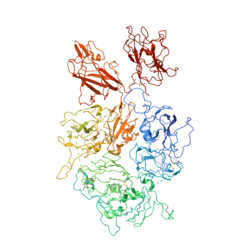
Structure of coagulation factor VIII bound to a patient-derived anti-C1 domain antibody inhibitor.

Childers, K.C.Avery, N.G.Estrada Alamo, K.A.Davulcu, O.Haynes, R.M.Lollar, P.Doering, C.B.Coxon, C.H.Spiegel, P.C.

(2023) Blood 

  • DOI: https://doi.org/10.1182/blood.2023020181
  • Primary Citation of Related Structures:  
    8G6I

  • PubMed Abstract: 
  • The development of pathogenic antibody inhibitors against coagulation factor VIII (FVIII) occurs in approximately 30% of congenital hemophilia A patients receiving FVIII replacement therapy as well as in all cases of acquired hemophilia A. KM33 is an anti-C1 domain antibody inhibitor previously isolated from a severe hemophilia A patient ...

    The development of pathogenic antibody inhibitors against coagulation factor VIII (FVIII) occurs in approximately 30% of congenital hemophilia A patients receiving FVIII replacement therapy as well as in all cases of acquired hemophilia A. KM33 is an anti-C1 domain antibody inhibitor previously isolated from a severe hemophilia A patient. In addition to potently blocking FVIII binding to von Willebrand factor and phospholipid surfaces, KM33 disrupts FVIII binding to lipoprotein receptor-related protein 1 (LRP1), which drives FVIII hepatic clearance and antigen presentation in dendritic cells. Here, we report on the structure of FVIII bound to NB33, a recombinant derivative of KM33, by single-particle cryo-electron microscopy. Structural analysis reveals the NB33 epitope localizes to FVIII residues R2090-S2094 and I2158-R2159 which constitute membrane-binding loops in the C1 domain. Further analysis reveals multiple FVIII lysine and arginine residues, previously shown to mediate binding to LRP1, dock onto an acidic cleft at the NB33 variable domain interface, thus blocking a putative LRP1 binding site. Together, these results demonstrate a novel mechanism of FVIII inhibition by a patient-derived antibody inhibitor and provide structural evidence toward engineering FVIII with reduced LRP1-mediated clearance.


    Organizational Affiliation

    Western Washington University, Bellingham, Washington, United States.


Macromolecules
Find similar proteins by:  (by identity cutoff)  |  3D Structure
Entity ID: 1
MoleculeChainsSequence LengthOrganismDetailsImage
Coagulation factor VIII chimera from human and pigA1,467Sus scrofaHomo sapiensMutation(s): 0 
Gene Names: F8CF8F8C
UniProt & NIH Common Fund Data Resources
Find proteins for P12263 (Sus scrofa)
Explore P12263 
Go to UniProtKB:  P12263
Find proteins for P00451 (Homo sapiens)
Explore P00451 
Go to UniProtKB:  P00451
PHAROS:  P00451
Entity Groups  
Sequence Clusters30% Identity50% Identity70% Identity90% Identity95% Identity100% Identity
UniProt GroupsP12263P00451
Protein Feature View
Expand
  • Reference Sequence
Find similar proteins by:  (by identity cutoff)  |  3D Structure
Entity ID: 2
MoleculeChainsSequence LengthOrganismDetailsImage
NB33 light chainB211Homo sapiensMutation(s): 0 
Entity Groups  
Sequence Clusters30% Identity50% Identity70% Identity90% Identity95% Identity100% Identity
Protein Feature View
Expand
  • Reference Sequence
Find similar proteins by:  (by identity cutoff)  |  3D Structure
Entity ID: 3
MoleculeChainsSequence LengthOrganismDetailsImage
NB33 heavy chainC215Homo sapiensMutation(s): 0 
Entity Groups  
Sequence Clusters30% Identity50% Identity70% Identity90% Identity95% Identity100% Identity
Protein Feature View
Expand
  • Reference Sequence
Oligosaccharides

Help

Entity ID: 4
MoleculeChainsChain Length2D DiagramGlycosylation3D Interactions
2-acetamido-2-deoxy-beta-D-glucopyranose-(1-4)-2-acetamido-2-deoxy-beta-D-glucopyranoseD, E 2N-Glycosylation Oligosaccharides Interaction
Experimental Data & Validation

Experimental Data

  • Method: ELECTRON MICROSCOPY
  • Resolution: 4.23 Å
  • Aggregation State: PARTICLE 
  • Reconstruction Method: SINGLE PARTICLE 
EM Software:
TaskSoftware PackageVersion
RECONSTRUCTIONcryoSPARC3.3.2
MODEL REFINEMENTPHENIX1.20

Structure Validation

View Full Validation Report


Entry History & Funding Information

Deposition Data


Funding OrganizationLocationGrant Number
National Institutes of Health/National Heart, Lung, and Blood Institute (NIH/NHLBI)United StatesR15HL135658
National Institutes of Health/National Heart, Lung, and Blood Institute (NIH/NHLBI)United StatesU54HL141981
National Institutes of Health/National Heart, Lung, and Blood Institute (NIH/NHLBI)United StatesR44HL117511
National Institutes of Health/National Heart, Lung, and Blood Institute (NIH/NHLBI)United StatesR44HL110448
National Institutes of Health/National Institute of General Medical Sciences (NIH/NIGMS)United StatesU24GM129547

Revision History  (Full details and data files)

  • Version 1.0: 2023-05-24
    Type: Initial release