Expression, purification and crystallization of N-acetyl-(R)-beta-phenylalanine acylases derived from Burkholderia sp. AJ110349 and Variovorax sp. AJ110348 and structure determination of the Burkholderia enzyme.
Kato, Y., Kawasaki, H., Nakamatsu, T., Matsuda, N., Natsume, R.(2023) Acta Crystallogr F Struct Biol Commun 79: 70-78
- PubMed: 36862095 Search on PubMedSearch on PubMed Central
- DOI: https://doi.org/10.1107/S2053230X23000730
- Primary Citation Related Structures:
8HUY, 8I59, 8I5A - PubMed Abstract:
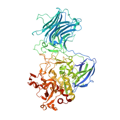
N-Acetyl-(R)-β-phenylalanine acylase is an enzyme that hydrolyzes the amide bond of N-acetyl-(R)-β-phenylalanine to produce enantiopure (R)-β-phenylalanine. In previous studies, Burkholderia sp. AJ110349 and Variovorax sp. AJ110348 were isolated as (R)-enantiomer-specific N-acetyl-(R)-β-phenylalanine acylase-producing organisms and the properties of the native enzyme from Burkholderia sp. AJ110349 were characterized. In this study, structural analyses were carried out in order to investigate the structure-function relationships of the enzymes derived from both organisms. The recombinant N-acetyl-(R)-β-phenylalanine acylases were crystallized by the hanging-drop vapor-diffusion method under multiple crystallization solution conditions. The crystals of the Burkholderia enzyme belonged to space group P4 1 2 1 2, with unit-cell parameters a = b = 112.70-112.97, c = 341.50-343.32 Å, and were likely to contain two subunits in the asymmetric unit. The crystal structure was solved by the Se-SAD method, suggesting that two subunits in the asymmetric unit form a dimer. Each subunit was composed of three domains, and they showed structural similarity to the corresponding domains of the large subunit of N,N-dimethylformamidase from Paracoccus sp. strain DMF. The crystals of the Variovorax enzyme grew as twinned crystals and were not suitable for structure determination. Using size-exclusion chromatography with online static light-scattering analysis, the N-acetyl-(R)-β-phenylalanine acylases were clarified to be dimeric in solution.
- Graduate School of Advanced Science and Technology, Tokyo Denki University, 5 Senju-asahi-cho, Adachi-ku, Tokyo 120-8551, Japan.
Organizational Affiliation: