8K2K

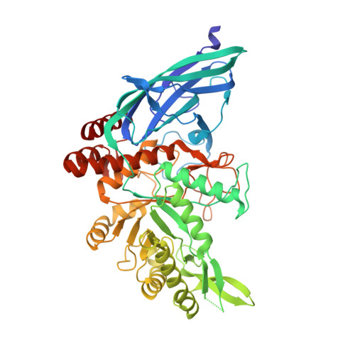
Crystal structure of Group 3 Oligosaccharide/Monosaccharide-releasing beta-N-acetylgalactosaminidase NgaDssm in complex with GalNAc-thiazoline

  • Classification: HYDROLASE
  • Organism(s): metagenome
  • Expression System: Escherichia coli
  • Mutation(s): No 

  • Deposited: 2023-07-12 Released: 2024-04-24 
  • Deposition Author(s): Sumida, T., Fushinobu, S.
  • Funding Organization(s): Japan Society for the Promotion of Science (JSPS), Mizutani Foundation for Glycoscience

Experimental Data Snapshot

  • Method: X-RAY DIFFRACTION
  • Resolution: 1.75 Å
  • R-Value Free: 0.206 
  • R-Value Work: 0.176 
  • R-Value Observed: 0.177 

Starting Model: in silico
View more details

wwPDB Validation   3D Report Full Report


Ligand Structure Quality Assessment 


This is version 1.1 of the entry. See complete history


Literature

Genetic and functional diversity of beta-N-acetylgalactosamine-targeting glycosidases expanded by deep-sea metagenome analysis.

Sumida, T.Hiraoka, S.Usui, K.Ishiwata, A.Sengoku, T.Stubbs, K.A.Tanaka, K.Deguchi, S.Fushinobu, S.Nunoura, T.

(2024) Nat Commun 15: 3543-3543

  • DOI: https://doi.org/10.1038/s41467-024-47653-2
  • Primary Citation of Related Structures:  
    8K2F, 8K2G, 8K2H, 8K2I, 8K2J, 8K2K, 8K2L, 8K2M, 8K2N

  • PubMed Abstract: 

    β-N-Acetylgalactosamine-containing glycans play essential roles in several biological processes, including cell adhesion, signal transduction, and immune responses. β-N-Acetylgalactosaminidases hydrolyze β-N-acetylgalactosamine linkages of various glycoconjugates. However, their biological significance remains ambiguous, primarily because only one type of enzyme, exo-β-N-acetylgalactosaminidases that specifically act on β-N-acetylgalactosamine residues, has been documented to date. In this study, we identify four groups distributed among all three domains of life and characterize eight β-N-acetylgalactosaminidases and β-N-acetylhexosaminidase through sequence-based screening of deep-sea metagenomes and subsequent searching of public protein databases. Despite low sequence similarity, the crystal structures of these enzymes demonstrate that all enzymes share a prototype structure and have diversified their substrate specificities (oligosaccharide-releasing, oligosaccharide/monosaccharide-releasing, and monosaccharide-releasing) through the accumulation of mutations and insertional amino acid sequences. The diverse β-N-acetylgalactosaminidases reported in this study could facilitate the comprehension of their structures and functions and present evolutionary pathways for expanding their substrate specificity.


  • Organizational Affiliation

    Research Center for Bioscience and Nanoscience, Japan Agency for Marine-Earth Science and Technology (JAMSTEC), Yokosuka, Japan. sumidat@jamstec.go.jp.


Macromolecules
Find similar proteins by:  (by identity cutoff)  |  3D Structure
Entity ID: 1
MoleculeChains Sequence LengthOrganismDetailsImage
Oligosaccharide/Monosaccharide-releasing beta-N-acetylgalactosaminidase542metagenomeMutation(s): 0 
Entity Groups  
Sequence Clusters30% Identity50% Identity70% Identity90% Identity95% Identity100% Identity
Sequence Annotations
Expand
  • Reference Sequence
Small Molecules
Ligands 4 Unique
IDChains Name / Formula / InChI Key2D Diagram3D Interactions
GNL
Query on GNL

Download Ideal Coordinates CCD File 
B [auth A](3aR,5R,6R,7R,7aR)-5-(hydroxymethyl)-2-methyl-5,6,7,7a-tetrahydro-3aH-pyrano[3,2-d][1,3]thiazole-6,7-diol
C8 H13 N O4 S
DRHXTSWSUAJOJZ-JAJWTYFOSA-N
SO4
Query on SO4

Download Ideal Coordinates CCD File 
D [auth A],
E [auth A]
SULFATE ION
O4 S
QAOWNCQODCNURD-UHFFFAOYSA-L
GOL
Query on GOL

Download Ideal Coordinates CCD File 
C [auth A]GLYCEROL
C3 H8 O3
PEDCQBHIVMGVHV-UHFFFAOYSA-N
CL
Query on CL

Download Ideal Coordinates CCD File 
F [auth A]CHLORIDE ION
Cl
VEXZGXHMUGYJMC-UHFFFAOYSA-M
Experimental Data & Validation

Experimental Data

  • Method: X-RAY DIFFRACTION
  • Resolution: 1.75 Å
  • R-Value Free: 0.206 
  • R-Value Work: 0.176 
  • R-Value Observed: 0.177 
  • Space Group: P 43 21 2
Unit Cell:
Length ( Å )Angle ( ˚ )
a = 53.537α = 90
b = 53.537β = 90
c = 428.292γ = 90
Software Package:
Software NamePurpose
REFMACrefinement
XDSdata reduction
Aimlessdata scaling
PHASERphasing

Structure Validation

View Full Validation Report


Ligand Structure Quality Assessment 


Entry History & Funding Information

Deposition Data


Funding OrganizationLocationGrant Number
Japan Society for the Promotion of Science (JSPS)JapanJP22K05398
Japan Society for the Promotion of Science (JSPS)JapanJP20K15444
Mizutani Foundation for GlycoscienceJapanFT100100291

Revision History  (Full details and data files)

  • Version 1.0: 2024-04-24
    Type: Initial release
  • Version 1.1: 2024-05-22
    Changes: Database references