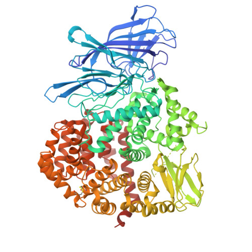
8P0I

Crystal structure of the open conformation of insulin-regulated aminopeptidase in complex with a small-MW inhibitor


Experimental Data Snapshot

  • Method: X-RAY DIFFRACTION
  • Resolution: 3.50 Å
  • R-Value Free: 0.277 
  • R-Value Work: 0.240 

Starting Model: experimental
View more details

wwPDB Validation   3D Report Full Report


Ligand Structure Quality Assessment 


This is version 1.1 of the entry. See complete history


Literature

Stabilization of the open conformation omicron f insulin-regulated aminopeptidase by a novel substrate-selective small-molecule inhibitor.

Mpakali, A.Georgaki, G.Buson, A.Findlay, A.D.Foot, J.S.Mauvais, F.X.van Endert, P.Giastas, P.Hamprecht, D.W.Stratikos, E.

(2024) Protein Sci 33: e5151-e5151

  • DOI: https://doi.org/10.1002/pro.5151
  • Primary Citation of Related Structures:  
    8P0I

  • PubMed Abstract: 

    Insulin-regulated aminopeptidase (IRAP) is an enzyme with important biological functions and the target of drug-discovery efforts. We combined in silico screening with a medicinal chemistry optimization campaign to discover a nanomolar inhibitor of IRAP based on a pyrazolylpyrimidine scaffold. This compound displays an excellent selectivity profile versus homologous aminopeptidases, and kinetic analysis suggests it utilizes an uncompetitive mechanism of action when inhibiting the cleavage of a typical dipeptidic substrate. Surprisingly, the compound is a poor inhibitor of the processing of the physiological cyclic peptide substrate oxytocin and a 10mer antigenic epitope precursor but displays a biphasic inhibition profile for the trimming of a 9mer antigenic peptide. While the compound reduces IRAP-dependent cross-presentation of an 8mer epitope in a cellular assay, it fails to block in vitro trimming of select epitope precursors. To gain insight into the mechanism and basis of this unusual selectivity for this inhibitor, we solved the crystal structure of its complex with IRAP. The structure indicated direct zinc(II) engagement by the pyrazolylpyrimidine scaffold and revealed that the compound binds to an open conformation of the enzyme in a pose that should block the conformational transition to the enzymatically active closed conformation previously observed for other low-molecular-weight inhibitors. This compound constitutes the first IRAP inhibitor targeting the active site that utilizes a conformation-specific mechanism of action, provides insight into the intricacies of the IRAP catalytic cycle, and highlights a novel approach to regulating IRAP activity by blocking its conformational rearrangements.


  • Organizational Affiliation

    National Centre for Scientific Research Demokritos, Athens, Greece.


Macromolecules
Find similar proteins by:  (by identity cutoff)  |  3D Structure
Entity ID: 1
MoleculeChains Sequence LengthOrganismDetailsImage
Leucyl-cystinyl aminopeptidase, pregnancy serum form
A, B
871Homo sapiensMutation(s): 0 
Gene Names: LNPEPOTASE
EC: 3.4.11.3
UniProt & NIH Common Fund Data Resources
Find proteins for Q9UIQ6 (Homo sapiens)
Explore Q9UIQ6 
Go to UniProtKB:  Q9UIQ6
PHAROS:  Q9UIQ6
GTEx:  ENSG00000113441 
Entity Groups  
Sequence Clusters30% Identity50% Identity70% Identity90% Identity95% Identity100% Identity
UniProt GroupQ9UIQ6
Glycosylation
Glycosylation Sites: 13Go to GlyGen: Q9UIQ6-1
Sequence Annotations
Expand
  • Reference Sequence
Oligosaccharides

Help

Entity ID: 2
MoleculeChains Length2D Diagram Glycosylation3D Interactions
beta-D-mannopyranose-(1-4)-2-acetamido-2-deoxy-beta-D-glucopyranose-(1-4)-2-acetamido-2-deoxy-beta-D-glucopyranose
C, E, G, I
3N-Glycosylation
Glycosylation Resources
GlyTouCan:  G15407YE
GlyCosmos:  G15407YE
GlyGen:  G15407YE
Entity ID: 3
MoleculeChains Length2D Diagram Glycosylation3D Interactions
alpha-D-mannopyranose-(1-3)-beta-D-mannopyranose-(1-4)-2-acetamido-2-deoxy-beta-D-glucopyranose-(1-4)-2-acetamido-2-deoxy-beta-D-glucopyranose
D, F
4N-Glycosylation
Glycosylation Resources
GlyTouCan:  G81315DD
GlyCosmos:  G81315DD
GlyGen:  G81315DD
Entity ID: 4
MoleculeChains Length2D Diagram Glycosylation3D Interactions
2-acetamido-2-deoxy-beta-D-glucopyranose-(1-4)-2-acetamido-2-deoxy-beta-D-glucopyranose
H, J, K, L
2N-Glycosylation
Glycosylation Resources
GlyTouCan:  G42666HT
GlyCosmos:  G42666HT
GlyGen:  G42666HT
Small Molecules
Ligands 3 Unique
IDChains Name / Formula / InChI Key2D Diagram3D Interactions
WC3 (Subject of Investigation/LOI)
Query on WC3

Download Ideal Coordinates CCD File 
BA [auth B],
S [auth A]
2-[2-(3,5-dimethylpyrazol-1-yl)-6-(4-methoxyphenyl)pyrimidin-4-yl]oxy-N-methyl-N-[(2-methylpyridin-3-yl)methyl]ethanamide
C26 H28 N6 O3
VJEOBRGFUNEYOH-UHFFFAOYSA-N
NAG
Query on NAG

Download Ideal Coordinates CCD File 
AA [auth B]
M [auth A]
N [auth A]
O [auth A]
P [auth A]
AA [auth B],
M [auth A],
N [auth A],
O [auth A],
P [auth A],
Q [auth A],
R [auth A],
U [auth A],
V [auth B],
W [auth B],
X [auth B],
Y [auth B],
Z [auth B]
2-acetamido-2-deoxy-beta-D-glucopyranose
C8 H15 N O6
OVRNDRQMDRJTHS-FMDGEEDCSA-N
ZN
Query on ZN

Download Ideal Coordinates CCD File 
CA [auth B],
T [auth A]
ZINC ION
Zn
PTFCDOFLOPIGGS-UHFFFAOYSA-N
Experimental Data & Validation

Experimental Data

  • Method: X-RAY DIFFRACTION
  • Resolution: 3.50 Å
  • R-Value Free: 0.277 
  • R-Value Work: 0.240 
  • Space Group: P 1 21 1
Unit Cell:
Length ( Å )Angle ( ˚ )
a = 68.229α = 90
b = 255.121β = 110
c = 73.274γ = 90
Software Package:
Software NamePurpose
PHENIXrefinement
PHENIXrefinement
XDSdata reduction
SCALAdata scaling
PHASERphasing

Structure Validation

View Full Validation Report


Ligand Structure Quality Assessment 


Entry History & Funding Information

Deposition Data


Funding OrganizationLocationGrant Number
European Union (EU)European Union--

Revision History  (Full details and data files)

  • Version 1.0: 2024-09-04
    Type: Initial release
  • Version 1.1: 2024-11-06
    Changes: Structure summary