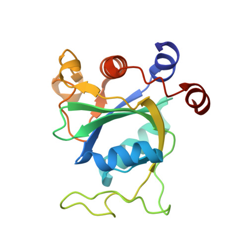
8Q2X

Crystal structure of YTHDC1 in complex with Compound 10 (ZA_294)


Experimental Data Snapshot

  • Method: X-RAY DIFFRACTION
  • Resolution: 1.60 Å
  • R-Value Free: 0.245 
  • R-Value Work: 0.211 
  • R-Value Observed: 0.212 

Starting Model: experimental
View more details

wwPDB Validation   3D Report Full Report


Ligand Structure Quality Assessment 


This is version 1.1 of the entry. See complete history


Literature

Structure-Based Design of a Potent and Selective YTHDC1 Ligand.

Zalesak, F.Nai, F.Herok, M.Bochenkova, E.Bedi, R.K.Li, Y.Errani, F.Caflisch, A.

(2024) J Med Chem 67: 9516-9535

  • DOI: https://doi.org/10.1021/acs.jmedchem.4c00599
  • Primary Citation of Related Structures:  
    8Q2Q, 8Q2R, 8Q2S, 8Q2T, 8Q2U, 8Q2V, 8Q2W, 8Q2X, 8Q2Y, 8Q31, 8Q32, 8Q33, 8Q35, 8Q37, 8Q38, 8Q39, 8Q3A, 8Q3G, 8Q4M, 8Q4N, 8Q4P, 8Q4Q, 8Q4R, 8Q4T, 8Q4U, 8Q4V, 8Q4W

  • PubMed Abstract: 

    N 6 -Adenosine methylation (m 6 A) is a prevalent post-transcriptional modification of mRNA, with YTHDC1 being the reader protein responsible for recognizing this modification in the cell nucleus. Here, we present a protein structure-based medicinal chemistry campaign that resulted in the YTHDC1 inhibitor 40 , which shows an equilibrium dissociation constant ( K d ) of 49 nM. The crystal structure of the complex (1.6 Å resolution) validated the design. Compound 40 is selective against the cytoplasmic m 6 A-RNA readers YTHDF1-3 and YTHDC2 and shows antiproliferative activity against the acute myeloid leukemia (AML) cell lines THP-1, MOLM-13, and NOMO-1. For the series of compounds that culminated into ligand 40 , the good correlation between the affinity in the biochemical assay and antiproliferative activity in the THP-1 cell line provides evidence of YTHDC1 target engagement in the cell. The binding to YTHDC1 in the cell is further supported by the cellular thermal shift assay. Thus, ligand 40 is a tool compound for studying the role of YTHDC1 in AML.


  • Organizational Affiliation

    Department of Biochemistry, University of Zurich, Winterthurerstrasse 190, CH-8057 Zurich, Switzerland.


Macromolecules
Find similar proteins by:  (by identity cutoff)  |  3D Structure
Entity ID: 1
MoleculeChains Sequence LengthOrganismDetailsImage
YTH domain-containing protein 1
A, B
166Homo sapiensMutation(s): 0 
Gene Names: YTHDC1KIAA1966YT521
UniProt & NIH Common Fund Data Resources
Find proteins for Q96MU7 (Homo sapiens)
Explore Q96MU7 
Go to UniProtKB:  Q96MU7
PHAROS:  Q96MU7
GTEx:  ENSG00000083896 
Entity Groups  
Sequence Clusters30% Identity50% Identity70% Identity90% Identity95% Identity100% Identity
UniProt GroupQ96MU7
Sequence Annotations
Expand
  • Reference Sequence
Experimental Data & Validation

Experimental Data

  • Method: X-RAY DIFFRACTION
  • Resolution: 1.60 Å
  • R-Value Free: 0.245 
  • R-Value Work: 0.211 
  • R-Value Observed: 0.212 
  • Space Group: P 1 21 1
Unit Cell:
Length ( Å )Angle ( ˚ )
a = 39.71α = 90
b = 103.45β = 105.81
c = 42.12γ = 90
Software Package:
Software NamePurpose
XDSdata reduction
XSCALEdata scaling
PHASERphasing
PHENIXrefinement

Structure Validation

View Full Validation Report


Ligand Structure Quality Assessment 


Entry History & Funding Information

Deposition Data


Funding OrganizationLocationGrant Number
Swiss National Science FoundationSwitzerland310030B_189363

Revision History  (Full details and data files)

  • Version 1.0: 2023-12-06
    Type: Initial release
  • Version 1.1: 2024-06-19
    Changes: Database references