8QCU

Lysozyme covalently bound to fac-[Re(CO)3-imidazole] complex, incubated for 38 weeks.


Experimental Data Snapshot

  • Method: X-RAY DIFFRACTION
  • Resolution: 1.15 Å
  • R-Value Free: 0.178 
  • R-Value Work: 0.159 
  • R-Value Observed: 0.160 

wwPDB Validation   3D Report Full Report


Ligand Structure Quality Assessment 


This is version 1.1 of the entry. See complete history


Literature

Time-series analysis of rhenium(I) organometallic covalent binding to a model protein for drug development.

Jacobs, F.J.F.Helliwell, J.R.Brink, A.

(2024) IUCrJ 11: 359-373

  • DOI: https://doi.org/10.1107/S2052252524002598
  • Primary Citation of Related Structures:  
    8QCU

  • PubMed Abstract: 

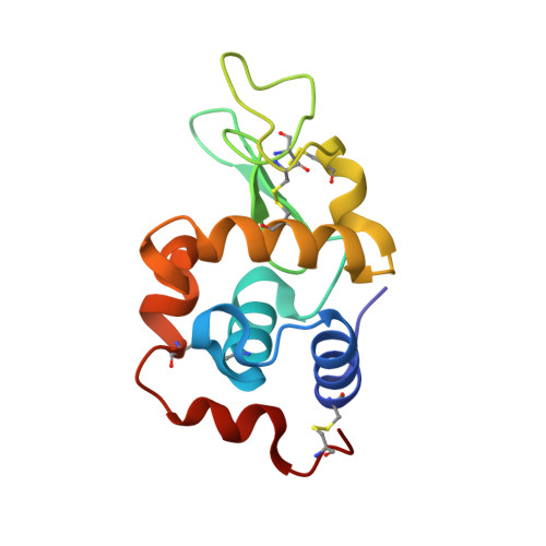
    Metal-based complexes with their unique chemical properties, including multiple oxidation states, radio-nuclear capabilities and various coordination geometries yield value as potential pharmaceuticals. Understanding the interactions between metals and biological systems will prove key for site-specific coordination of new metal-based lead compounds. This study merges the concepts of target coordination with fragment-based drug methodologies, supported by varying the anomalous scattering of rhenium along with infrared spectroscopy, and has identified rhenium metal sites bound covalently with two amino acid types within the model protein. A time-based series of lysozyme-rhenium-imidazole (HEWL-Re-Imi) crystals was analysed systematically over a span of 38 weeks. The main rhenium covalent coordination is observed at His15, Asp101 and Asp119. Weak (i.e. noncovalent) interactions are observed at other aspartic, asparagine, proline, tyrosine and tryptophan side chains. Detailed bond distance comparisons, including precision estimates, are reported, utilizing the diffraction precision index supplemented with small-molecule data from the Cambridge Structural Database. Key findings include changes in the protein structure induced at the rhenium metal binding site, not observed in similar metal-free structures. The binding sites are typically found along the solvent-channel-accessible protein surface. The three primary covalent metal binding sites are consistent throughout the time series, whereas binding to neighbouring amino acid residues changes through the time series. Co-crystallization was used, consistently yielding crystals four days after setup. After crystal formation, soaking of the compound into the crystal over 38 weeks is continued and explains these structural adjustments. It is the covalent bond stability at the three sites, their proximity to the solvent channel and the movement of residues to accommodate the metal that are important, and may prove useful for future radiopharmaceutical development including target modification.


  • Organizational Affiliation

    Department of Chemistry, University of the Free State, Nelson Mandela Drive, Bloemfontein, 9301, South Africa.


Macromolecules
Find similar proteins by:  (by identity cutoff)  |  3D Structure
Entity ID: 1
MoleculeChains Sequence LengthOrganismDetailsImage
Lysozyme C129Gallus gallusMutation(s): 0 
EC: 3.2.1.17
UniProt
Find proteins for P00698 (Gallus gallus)
Explore P00698 
Go to UniProtKB:  P00698
Entity Groups  
Sequence Clusters30% Identity50% Identity70% Identity90% Identity95% Identity100% Identity
UniProt GroupP00698
Sequence Annotations
Expand
  • Reference Sequence
Small Molecules
Ligands 7 Unique
IDChains Name / Formula / InChI Key2D Diagram3D Interactions
VHL (Subject of Investigation/LOI)
Query on VHL

Download Ideal Coordinates CCD File 
G [auth A],
I [auth A]
Tricarbonyl di(imidazole) rhenium(I)
C9 H6 N4 O3 Re
MWUHNOZKPZGOSW-UHFFFAOYSA-N
REI (Subject of Investigation/LOI)
Query on REI

Download Ideal Coordinates CCD File 
H [auth A]Tricarbonyl (aqua) (imidazole) rhenium(I)
C6 H6 N2 O4 Re
KPOHHEXTCRHHSE-UHFFFAOYSA-N
RE
Query on RE

Download Ideal Coordinates CCD File 
K [auth A]
L [auth A]
M [auth A]
N [auth A]
O [auth A]
K [auth A],
L [auth A],
M [auth A],
N [auth A],
O [auth A],
P [auth A]
RHENIUM
Re
WUAPFZMCVAUBPE-UHFFFAOYSA-N
BR
Query on BR

Download Ideal Coordinates CCD File 
R [auth A]BROMIDE ION
Br
CPELXLSAUQHCOX-UHFFFAOYSA-M
IMD
Query on IMD

Download Ideal Coordinates CCD File 
J [auth A]IMIDAZOLE
C3 H5 N2
RAXXELZNTBOGNW-UHFFFAOYSA-O
CL
Query on CL

Download Ideal Coordinates CCD File 
B [auth A],
C [auth A],
D [auth A],
E [auth A],
Q [auth A]
CHLORIDE ION
Cl
VEXZGXHMUGYJMC-UHFFFAOYSA-M
NA
Query on NA

Download Ideal Coordinates CCD File 
F [auth A]SODIUM ION
Na
FKNQFGJONOIPTF-UHFFFAOYSA-N
Experimental Data & Validation

Experimental Data

  • Method: X-RAY DIFFRACTION
  • Resolution: 1.15 Å
  • R-Value Free: 0.178 
  • R-Value Work: 0.159 
  • R-Value Observed: 0.160 
  • Space Group: P 43 21 2
Unit Cell:
Length ( Å )Angle ( ˚ )
a = 81.12α = 90
b = 81.12β = 90
c = 37.19γ = 90
Software Package:
Software NamePurpose
PHENIXrefinement
xia2data reduction
xia2data scaling
PHASERphasing

Structure Validation

View Full Validation Report


Ligand Structure Quality Assessment 


Entry History & Funding Information

Deposition Data


Funding OrganizationLocationGrant Number
National Research Foundation in South AfricaSouth Africa137759
Other privateSASOL University Collaboration Initiative
Other governmentDHET Future Professors Program
Other governmentSTART program: BAG MX28402-9, BAG mx28402-14, and BAG mx28402-19

Revision History  (Full details and data files)

  • Version 1.0: 2024-05-01
    Type: Initial release
  • Version 1.1: 2024-05-15
    Changes: Database references