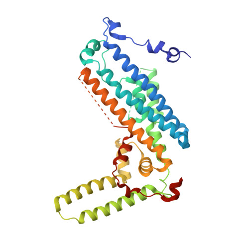
8R5R

Structure of apo TDO with a bound inhibitor


Experimental Data Snapshot

  • Method: X-RAY DIFFRACTION
  • Resolution: 3.08 Å
  • R-Value Free: 0.252 
  • R-Value Work: 0.227 
  • R-Value Observed: 0.228 

Starting Model: experimental
View more details

wwPDB Validation   3D Report Full Report


Ligand Structure Quality Assessment 


This is version 1.1 of the entry. See complete history


Literature

Discovery and binding mode of small molecule inhibitors of the apo form of human TDO2.

Lotz-Jenne, C.Lange, R.Cren, S.Bourquin, G.Goglia, L.Kimmerlin, T.Wicki, M.Muller, M.Artico, N.Ackerknecht, S.Pfaff, P.Joesch, C.Mac Sweeney, A.

(2024) Sci Rep 14: 27937-27937

  • DOI: https://doi.org/10.1038/s41598-024-78981-4
  • Primary Citation of Related Structures:  
    8R5Q, 8R5R, 9EZJ

  • PubMed Abstract: 

    Tryptophan-2,3-dioxygenase (TDO2) and indoleamine-2,3-dioxygenase (IDO1) are structurally distinct heme enzymes that catalyze the conversion of L-tryptophan to N-formyl-kynurenine, and play important roles in metabolism, inflammation, and tumor immune surveillance. The enzymes can adopt an inactive, heme-free (apo) state or an active, heme-containing (holo) state, with the balance between them varying dynamically according to biological conditions. Inhibitors of holo-TDO2 are known but, despite several advantages of the heme-free state as a drug target, no inhibitors of apo-TDO2 have been reported. We describe the discovery of the first apo-TDO2 binding inhibitors, to our knowledge, and their inhibition of cellular TDO2 activity at low nanomolar concentrations. The crystal structure of a potent, small molecule inhibitor bound to apo-TDO2 reveals its detailed binding interactions within the large, hydrophobic heme binding pocket of the active site.


  • Organizational Affiliation

    Drug discovery, Idorsia Pharmaceuticals Limited, Hegenheimermattweg 91, Allschwil, Basel-Land, 4123, Switzerland. carina.lotz@idorsia.com.


Macromolecules
Find similar proteins by:  (by identity cutoff)  |  3D Structure
Entity ID: 1
MoleculeChains Sequence LengthOrganismDetailsImage
Tryptophan 2,3-dioxygenase
A, B, C, D
358Homo sapiensMutation(s): 0 
Gene Names: TDO2
EC: 1.13.11.11
UniProt & NIH Common Fund Data Resources
Find proteins for P48775 (Homo sapiens)
Explore P48775 
Go to UniProtKB:  P48775
PHAROS:  P48775
GTEx:  ENSG00000151790 
Entity Groups  
Sequence Clusters30% Identity50% Identity70% Identity90% Identity95% Identity100% Identity
UniProt GroupP48775
Sequence Annotations
Expand
  • Reference Sequence
Small Molecules
Ligands 2 Unique
IDChains Name / Formula / InChI Key2D Diagram3D Interactions
Y5N (Subject of Investigation/LOI)
Query on Y5N

Download Ideal Coordinates CCD File 
E [auth A],
G [auth B],
I [auth C],
K [auth D]
3-chloranyl-~{N}-[(1~{S})-1-(6-chloranylpyridin-3-yl)-2-phenyl-ethyl]aniline
C19 H16 Cl2 N2
FPQMXEJXTNDYME-SFHVURJKSA-N
ZIQ
Query on ZIQ

Download Ideal Coordinates CCD File 
F [auth A],
H [auth B],
J [auth C],
L [auth D]
alpha-methyl-L-tryptophan
C12 H14 N2 O2
ZTTWHZHBPDYSQB-LBPRGKRZSA-N
Experimental Data & Validation

Experimental Data

Unit Cell:
Length ( Å )Angle ( ˚ )
a = 91.738α = 90
b = 132.906β = 90
c = 137.495γ = 90
Software Package:
Software NamePurpose
BUSTERrefinement
autoPROCdata reduction
autoPROCdata scaling
PHASERphasing

Structure Validation

View Full Validation Report


Ligand Structure Quality Assessment 


Entry History & Funding Information

Deposition Data


Funding OrganizationLocationGrant Number
Not funded--

Revision History  (Full details and data files)

  • Version 1.0: 2024-01-17
    Type: Initial release
  • Version 1.1: 2024-11-27
    Changes: Database references, Structure summary