Fragment-Based Development of Small Molecule Inhibitors Targeting Mycobacterium tuberculosis Cholesterol Metabolism.
Kavanagh, M.E., McLean, K.J., Gilbert, S.H., Amadi, C.N., Snee, M., Tunnicliffe, R.B., Arora, K., Boshoff, H.I.M., Fanourakis, A., Rebollo-Lopez, M.J., Ortega, F., Levy, C.W., Munro, A.W., Leys, D., Abell, C., Coyne, A.G.(2025) J Med Chem 68: 14416-14441
- PubMed: 40653654 Search on PubMed
- DOI: https://doi.org/10.1021/acs.jmedchem.5c00478
- Primary Citation Related Structures:
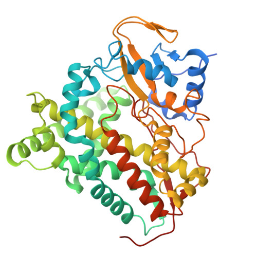
7P5T, 7QQ7, 7ZGL, 7ZIC, 8S4M, 8S53 - PubMed Abstract:
Tuberculosis is the deadliest infectious disease in history and new drugs are urgently required to combat multidrug-resistant (MDR) strains of Mycobacterium tuberculosis ( Mtb ). Here, we exploit the relience of Mtb on host-derived cholesterol to develop a novel class of antitubercular compounds that target Mtb CYP125 and CYP142; the enzymes that catalyze the first step of cholesterol metabolism. A combination of fragment screening and structure-based drug design was used to identify a hit compound and guide synthetic optimization of a dual CYP125/142 ligand 5m ( K D 40-160 nM), which potently inhibits enzyme activity in vitro ( K I < 100 nM), and the growth of Mtb in extracellular (MIC 99 0.4-1.5 μM) and intracellular assays (IC 50 1.7 μM). The structural data and lead compounds reported here will help study Mtb cholesterol metabolism and guide the development of novel antibiotics to combat MDR Mtb.
- Yusuf Hamied Department of Chemistry, University of Cambridge, Lensfield Road, Cambridge CB2 1EW, U.K.
Organizational Affiliation: