






 Download Mendeley
Crystal structure of Zn2+ bound calprotectin
Perera, Y.R., Nassif, A.R., Garcia, V., Guillen, R.M., Chazin, W.J.To be published.
Experimental Data Snapshot
Starting Model: experimental
View more details
wwPDB Validation 3D Report Full Report

Entity ID: 1 | |||||
|---|---|---|---|---|---|
| Molecule | Chains | Sequence Length | Organism | Details | Image |
| Protein S100-A8 | 88 | Homo sapiens | Mutation(s): 4 Gene Names: S100A8, CAGA, CFAG, MRP8 | 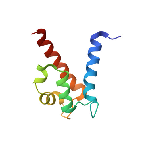 | |
UniProt & NIH Common Fund Data Resources | |||||
GTEx: ENSG00000143546 | |||||
Entity Groups | |||||
| Sequence Clusters | 30% Identity50% Identity70% Identity90% Identity95% Identity100% Identity | ||||
| UniProt Group | P05109 | ||||
Sequence AnnotationsExpand | |||||
Reference Sequence | |||||
Entity ID: 2 | |||||
|---|---|---|---|---|---|
| Molecule | Chains | Sequence Length | Organism | Details | Image |
| Protein S100-A9 | 99 | Homo sapiens | Mutation(s): 2 Gene Names: S100A9, CAGB, CFAG, MRP14 | 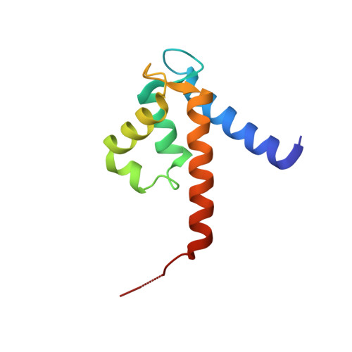 | |
UniProt & NIH Common Fund Data Resources | |||||
GTEx: ENSG00000163220 | |||||
Entity Groups | |||||
| Sequence Clusters | 30% Identity50% Identity70% Identity90% Identity95% Identity100% Identity | ||||
| UniProt Group | P06702 | ||||
Sequence AnnotationsExpand | |||||
Reference Sequence | |||||
| Ligands 3 Unique | |||||
|---|---|---|---|---|---|
| ID | Chains | Name / Formula / InChI Key | 2D Diagram | 3D Interactions | |
| 2PE Download:Ideal Coordinates CCD File | G [auth A], J [auth B] | NONAETHYLENE GLYCOL C18 H38 O10 YZUUTMGDONTGTN-UHFFFAOYSA-N | 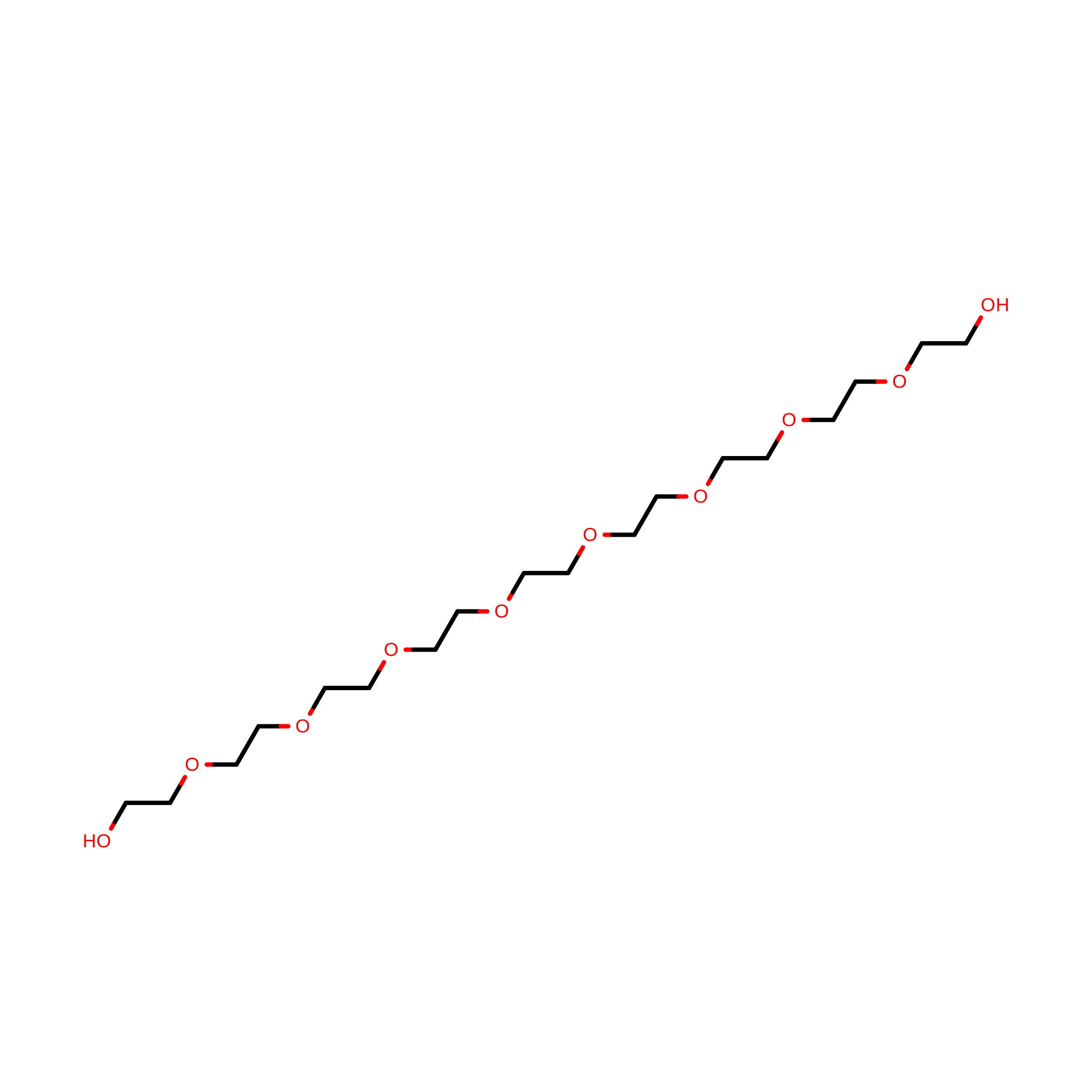 | ||
| ZN (Subject of Investigation/LOI) Download:Ideal Coordinates CCD File | K [auth C] | ZINC ION Zn PTFCDOFLOPIGGS-UHFFFAOYSA-N |  | ||
| CA Download:Ideal Coordinates CCD File | E [auth A] F [auth A] H [auth B] I [auth B] L [auth C] | CALCIUM ION Ca BHPQYMZQTOCNFJ-UHFFFAOYSA-N |  | ||
| Length ( Å ) | Angle ( ˚ ) |
|---|---|
| a = 58.436 | α = 90 |
| b = 76.935 | β = 90 |
| c = 106.793 | γ = 90 |
| Software Name | Purpose |
|---|---|
| PHENIX | refinement |
| Coot | model building |
| PHENIX | phasing |
| HKL-2000 | data reduction |
| XDS | data scaling |
| Funding Organization | Location | Grant Number |
|---|---|---|
| National Institutes of Health/National Institute of General Medical Sciences (NIH/NIGMS) | United States | R01 AI127793 |
| National Institutes of Health/Eunice Kennedy Shriver National Institute of Child Health & Human Development (NIH/NICHD) | United States | AI101171 |








